Key Highlights
  • Brain regions traditionally known as ‘non-eloquent’ often play important roles in high-level brain networks. Without more personalized information, it is challenging to map surgical corridors through them without causing deficits.
  • Recent literature has shown that ‘eloquent’ areas are often hubs for large-scale networks. However in some patients, these hubs appear in other regions of the brain.
  • Connectomics-based analysis can help neurosurgeons gain a more holistic perspective of brain network involvement in each case and is essential for moving beyond simplifying regions as being ‘eloquent’ and ‘non-eloquent’.

Eloquence and the neurosurgical trade-off

Brain tumor surgery aims to resect the maximal amount of tumor tissue while sacrificing the least amount of neurological function, a trade-off referred to as ‘onco-functional balance’1. Classic neurosurgery strategy emphasizes preserving ‘eloquent’ regions responsible for core brain functions, such as movement and speech, over so-called ‘non-eloquent’ regions that do not have an obvious function2.

However, while their function may not be immediately apparent, it does not mean they are sacrificial.

With growing connectomic research, it is increasingly clear that many ‘non-eloquent’ regions play crucial roles in higher-order brain networks. These networks underpin higher cognitive processes and are essential to preserving a patient’s functional independence.

Further, the core functions that often concern surgeons - motor, vision, and language - are rarely the manifestation of a single ‘eloquent’ region. Though eloquent regions are large contributors to these functions, they too are often part of broader networks which rely on a number of ‘non-eloquent’ regions to perform in a healthy and typical manner. Thus, even when eloquent regions are successfully avoided, these functions can still be impacted by damage to their network-constituent regions during surgery1.

Recognizing the above, advancing neurosurgery and improving patient outcomes means the adoption of more nuanced surgical planning techniques. These techniques will rely on the broader consideration of large scale brain networks, and increasing levels of personalization in preoperative planning practices3.

Major brain networks

Advances in neuroimaging and connectomics have shifted the focus away from individual cortical regions towards an understanding that brain function depends on large brain networks comprised of numerous parcels2. This work has identified major brain networks:

Sensorimotor network

Considered a priority during neurosurgery, the sensorimotor network is responsible for somatic sensation and motor planning and execution. The sensorimotor network is distributed over the primary and supplementary sensorimotor areas, cingulate cortex, dorsal premotor cortex, and the ventral premotor cortex.

Limbic system

The limbic system is one of the oldest brain networks and regulates vital brain functions like emotions, response to stressful stimuli, memory, and learning. This system is distributed across cortical and subcortical regions, including the hippocampus, amygdala, and several cortical areas in the temporal and orbitofrontal lobes.

The limbic system as seen in Quicktome

This image shows the location of a subject's limbic system, determined by Omniscient's personalized brain mapping platform.

Salience network

Spread across the frontal lobe, posterior cingulate cortex (PCC), insular cortex, and cingulate cortex, the salience network (SN) controls attentional state by deciding whether the default mode network or central executive network is active at a given time. It also processes sensorimotor information and is central to cognition.

Default mode network

The default mode network (DMN) can be thought of as the idle operating mode for the brain when it is not engaged with a task or planning. Brain regions such as the ACC, PCC, area 8Ad of the frontal lobe, and the lateral parietal lobe are components of the DMN.

Dorsal attention network

Sustained attention is made possible by the dorsal attention network (DAN), the aperture of the brain. Areas include the frontal lobe, parietal lobe, intraparietal sulcus, temporal lobe, and occipital lobe.

Central executive network

The central executive network (CEN) acts as a control network that engages in higher-level cognitive tasks and functions in tandem or anticorrelation with the other main networks. Numerous areas within the frontal lobe are associated with the CEN and regions in the parietal and temporal lobes.

Visual system

Visual processing is handled by the aptly named visual system. The visual system consists of the medial areas, such as V1, and 3 'streams' of information flow—the dorsal, ventral, and lateral streams.

How neurosurgery arrived at ‘eloquence’, and how to move on from it

Until the advent of modern neuroimaging and EEG techniques, discerning the function of brain regions was dependent on one premise - if the physiological processes of a region were disrupted, and a physical symptom were to present, then that region and that symptom were causally linked. Early on, this meant observing the symptoms of stroke patients and comparing regions of damaged brain with their physical symptoms. Over time, this developed into cortical stimulation techniques which could reproduce these symptoms temporarily through the application of electrical stimulation. For example, Broca's area could be identified by finding the region where stimulation causes speech arrest. Because physiological disruption could cause such profound physical responses in these regions, they were considered ‘eloquent’.

These cortical stimulation techniques remain a staple of neurosurgical theory and strategy. Indeed, its relevance is uncontroversial. Areas that contribute directly to core neurological functions should be preserved with utmost priority.

The implication, however, is that any region that does not cause an observable physical reaction when stimulated must be ‘non-eloquent’. Without additional anatomical information, it further implies all ‘non-eloquent’ regions are of equal level of unimportance. Thus, one resultant surgical strategy is to take the shortest possible surgical corridor. After all, if all non-eloquent regions are equally unimportant, then the objective would be to cut as little of it as possible.

With the substantial incidence of neurosurgical deficits, it is clear that this often does not lead to optimal patient outcomes and has become particularly challenged with the availability of new techniques to quantify a brain region’s contribution to neurological function.

Neuroimaging-based connectomics, for example, enables consideration of broader brain connectivity to quantify regions that do not respond to direct stimulation, and help separate the role and importance of one region from another.

This enables the surgeon to ask: amongst ‘non-eloquent’ brain tissue, is there a surgical corridor that has advantages over another?

Tractography GIF

Tractography is one way to visualize the brain’s ‘connectome’.

Quantifying the importance of brain regions using connectomics

The brain has an extraordinary capacity to re-organize itself if specific areas are damaged. These areas, termed the rich-club, are highly efficient hubs that integrate information from multiple other regions5,6. Additionally, Ahsan and colleagues (2020) reported that PageRank centrality, a measure of connectedness to other regions, was a better predictor than other measures at identifying brain areas relevant to neurosurgery2. Considering that several of these classically identified areas are centrally located in network topology, damage to seemingly ineloquent regions alters the inputs to these hubs in ways that may or may not translate to functional impairments1.

Connectomics also provides a method to assess damage caused to major brain networks. Work performed by Hart and colleagues (2016) characterized consistent alterations in connectivity between a set of hub regions in pre-op temporal-parietal-occipital glioblastoma7. Functional connectivity changes have also been noted in patients suffering from TBI, stroke, and even hypertension8-10.

Variation between individuals is apparent whether one operates based on eloquence or centrality; otherwise, there would be no need for cortical mapping of awake patients during surgery1. Networks, as one may expect, differ just as much as individual regions. A study of a large pool of diffusion neuroimaging data from healthy subjects by Yeung and colleagues revealed a subset of individuals with strong hub activity in unexpected regions11. Glioma patients are much more heterogeneous and display distinct connectivity profile differences that could help inform resection that minimizes further harm11-13.

One such technique in development, supra-maximal resection, relies on the identification of essential hub areas to maximize post-surgical cortical organization while resecting more tissue than what is usually resected using traditional approaches6. In addition, brain networks can be used to apply techniques such as deep brain stimulation (DBS) more precisely. Evidence suggests that DBS can cause widespread effects that propagate along white matter. Using a combination of fiber tractography, fMRI, and advanced diffusion imaging modalities, it may be possible to apply DBS in more nuanced ways that target the network alterations of an individual patient13.

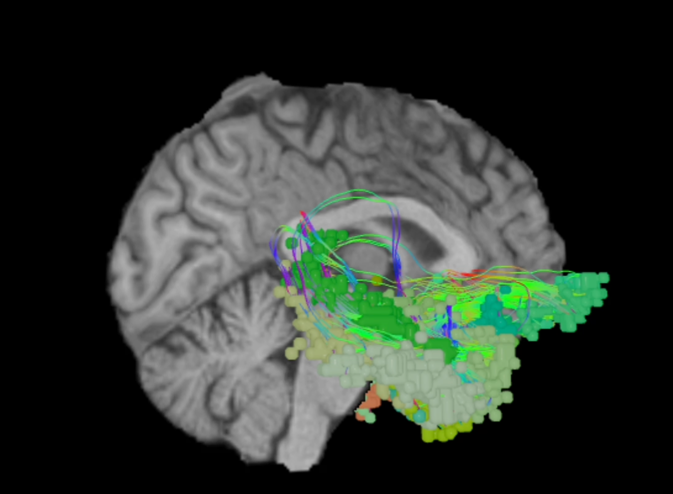
By mapping an individual patient's unique connectivity patterns, it may even be possible to predict functional deficits before surgery. In 2021, a team of researchers used Omniscient’s Quicktome software to see if the regions of strong hubness it identified could be correlated with neurological outcomes in 15 patients undergoing keyhole surgery12. Quicktome accurately predicted the functional deficits that emerged in seven of the eight patients with new post-operative impairments. The only outlier in this small study suffered a stroke shortly after surgery, potentially confounding results.

Quicktome was designed using machine-learning clustering techniques to rapidly analyze millions of data points from patient MRI scans based on connectivity profiles. This data is processed to generate an individualized map of a patient's brain networks. Quicktome has the added benefit of being cloud-based, so there is no need for new equipment as it seamlessly integrates into existing systems. Moreover, due to its cloud-based approach, surgeons are able to experience a hands-on, free trial of Quicktome by booking in a demo with their local representative.

Contact us now to get started or dig deeper into connectomics with our Connectome Academy.

References
Expand to see full list of references
  1. Dadario NB, Brahimaj B, Yeung J, Sughrue ME. Reducing the Cognitive Footprint of Brain Tumor Surgery. Front Neurol. 2021;12:711646.
  2. Ahsan SA, Chendeb K, Briggs RG, et al. Beyond eloquence and onto centrality: a new paradigm in planning supratentorial neurosurgery. J Neurooncol. 2020;146(2):229-238.
  3. Duffau H. Brain connectomics applied to oncological neuroscience: from a traditional surgical strategy focusing on glioma topography to a meta-network approach. Acta Neurochir (Wien). 2021;163(4):905-917.
  4. Yeo BT, Krienen FM, Sepulcre J, et al. The organization of the human cerebral cortex estimated by intrinsic functional connectivity. J Neurophysiol. 2011;106(3):1125-1165.
  5. Kim DJ, Min BK. Rich-club in the brain's macrostructure: Insights from graph theoretical analysis. Comput Struct Biotechnol J. 2020;18:1761-1773.
  6. Ius T, Angelini E, Thiebaut de Schotten M, Mandonnet E, Duffau H. Evidence for potentials and limitations of brain plasticity using an atlas of functional resectability of WHO grade II gliomas: towards a 'minimal common brain'. Neuroimage. 2011;56(3):992-1000.
  7. Hart MG, Price SJ, Suckling J. Connectome analysis for pre-operative brain mapping in neurosurgery. Br J Neurosurg. 2016;30(5):506-517.
  8. Carnevale L, Maffei A, Landolfi A, Grillea G, Carnevale D, Lembo G. Brain Functional Magnetic Resonance Imaging Highlights Altered Connections and Functional Networks in Patients With Hypertension. Hypertension. 2020;76(5):1480-1490.
  9. Dimitriadis SI, Zouridakis G, Rezaie R, Babajani-Feremi A, Papanicolaou AC. Functional connectivity changes detected with magnetoencephalography after mild traumatic brain injury. Neuroimage Clin. 2015;9:519-531.
  10. Li W, Li Y, Zhu W, Chen X. Changes in brain functional network connectivity after stroke. Neural Regen Res. 2014;9(1):51-60.
  11. Yeung JT, Taylor HM, Young IM, Nicholas PJ, Doyen S, Sughrue ME. Unexpected hubness: a proof-of-concept study of the human connectome using pagerank centrality and implications for intracerebral neurosurgery. J Neurooncol. 2021;151(2):249-256.
  12. Yeung JT, Taylor HM, Nicholas PJ, et al. Using Quicktome for Intracerebral Surgery: Early Retrospective Study and Proof of Concept. World Neurosurg. 2021;154:e734-e742.
  13. Henderson F, Abdullah KG, Verma R, Brem S. Tractography and the connectome in neurosurgical treatment of gliomas: the premise, the progress, and the potential. Neurosurg Focus. 2020;48(2):E6.