The human brain is an organ of billions of synapses firing in even greater numbers of patterns and sequences. These complex electrical processes are responsible for our every thought, emotion, function, and dysfunction. Understanding how the brain is connected and what these connections mean will drastically help us better understand mental health, cognition, and even what makes us human.

Of course, this is no small challenge. While scientists and doctors have poured over the brain’s structural and chemical properties for centuries, for the most part, the brain's connective properties have been overwhelming and poorly understood—at least by the human mind. As physicist Emerson M. Pugh put it, “If the brain were so simple that we could understand it, we’d be so simple that we couldn’t.”

Surmounting this certainly would sound like science fiction had it not been for the advent of big data, machine learning, and artificial intelligence. Today, challenges of complexity are ultimately challenges of data processing and the brain is no exception. In fact, it may just be the greatest—and most exciting—data challenge that we can solve.

… Enter Connectomics.

What is Connectomics?

Connectomics is the study of the connections in the nervous system. Connectomics explores how neurons in the brain and nervous system are connected and interact with each other to produce behavior and cognition. This includes both the structural connections between neurons, as well as the functional connections and communication between them.

The entire set of complex connections that make up the human brain is known as the brain’s connectome. The connectome is structurally connected by white matter between the cortex and subcortical structures (collectively known as the gray matter), and these connections act as wires to transfer information between functional areas of the brain. Think of all the individual connections in your computer carrying bits of data from different processing units - but many, many magnitudes more complex.

Due to its complexity, methods to analyze the connectome are liked to big data approaches. Non-invasive neuroimaging combined with highly complex statistical models are used to construct and decipher computer-generated maps of the brain's functional and structural connections.

As such, the word "connectomics" borrows the “-omics” suffix from “genomics,” which is the use of big data to study genetics in organisms. 

Visualizing the brain's connectome has become increasingly more sophisticated in both the lab and the operating room with non-invasive technologies such as tractography.

connectomics neurosurgery

Tractography is one way to visualize the brain’s ‘connectome’

A brief history of human brain mapping

In 1909, German neurologist, Korbinian Brodmann, detailed a brain parcellation based on anatomical and cellular structure of the brain’s surface1 - His model divided the human brain into around 47 parts, or ‘parcellations’, each with a supposed functional role. Area 4, for example, was responsible for motor functions, and Area 17 for visual processing. Brodmann did this by examining the cellular properties of the cerebral cortex, attempting to define boundaries where cellular structure changed. 

A century on, subsequent studies have validated many aspects of this early ‘brain map’. We know for example that damage to Area 4 will consistently cause deficits to movement and Area 17 to sight. Using similar techniques, there have even been several new areas that have been added or subdivided from existing areas.

human brain mapping

Broadmann brain mapping


The localizationist model of Brodmann’s original 47 areas.

The Brodmann model is, however, limited in explaining more complex neurological functions.

For starters, the method by which the Brodmann map identified functional areas of the brain was unlikely to be completely accurate - just like the examination of the earth’s surface alone does not meaningfully assist in drawing country, state, and city boundaries.

Indeed, it does not say much about how different areas of the brain interact, just as a world map does not provide much insight into geopolitics. Yet we know now that these interactions are responsible for important processes such as comprehension, emotion, and thinking. When these processes are compromised, they are also responsible for mental and neurological disorders such as depression, autism, and schizophrenia - all of which, as a result, have been poorly understood.

Limitations of the Brodmann ‘brain map’:
  • The physically observable differences between regions did not necessarily correlate with the functions they performed
  • The model grouped multiple regions that actually performed distinct, often unrelated functions
  • It does not to explain anything beyond very basic function such as movement and speech (and even then, did so simply)
  • Studied only the gray matter of the brain, and not the white matter that wired it together
  • Locations differed between different individuals - especially those with brain injury

However, while our technologies to help navigate street directions and to remain informed about world affairs have all grown drastically more powerful and accessible, most doctors and researchers have seen little changes to their tool kits to navigate and understand the brain.

limitations of Broadmann's brain map

Just as a world map alone cannot explain the complex interrelationships and interactions between countries and cultures, the Brodmann map does little to help us understand complex brain function and disorders.

The Human Connectome Project

In 2009, the Human Connectome Project (HCP) commenced a five-year effort to digitally map the structural and functional neural connections in the human brain. The $38.5M project began with a Blueprint Grand Challenge grant from the National Institute of Health and formed consortiums across top neuroscience institutions in the world including Washington University, Oxford, the University of Minnesota, Harvard, and UCLA.

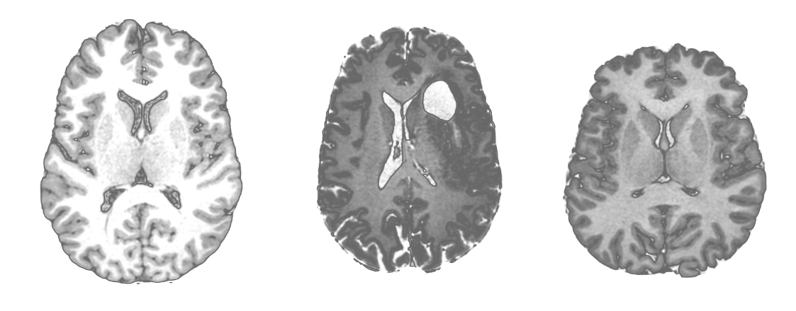
Collectively over a thousand subjects each underwent detailed studies of their brain using advanced, MRI-based techniques.

The HCP made substantial improvements in techniques to map the brain, and completely overhauled the alphabet and language used to study it. 

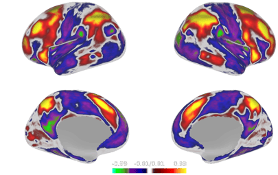
The result was a new brain map, the HCP Parcellation, or Atlas, that defined discrete brain areas based on their functional roles and how they were both functionally and structurally connected. This included 83 areas from previous studies and 97 that were previously unknown, totaling 180 in each hemisphere (or 360 in total)2.

Notably, the detail offered by this brain map was sufficiently intricate to explain higher brain function, yet still ‘simple’ enough to be analyzed by neuroscientists and (more importantly) computers and AI3.

The HCP parcellation differs from Brodmann’s because it is a multimodal method of identifying functional areas and their connections. Rather than looking at anatomical structure alone, the HCP atlas segments the brain based on cortical architecture, function, functional connectivity, and/or topography.

human connectome project

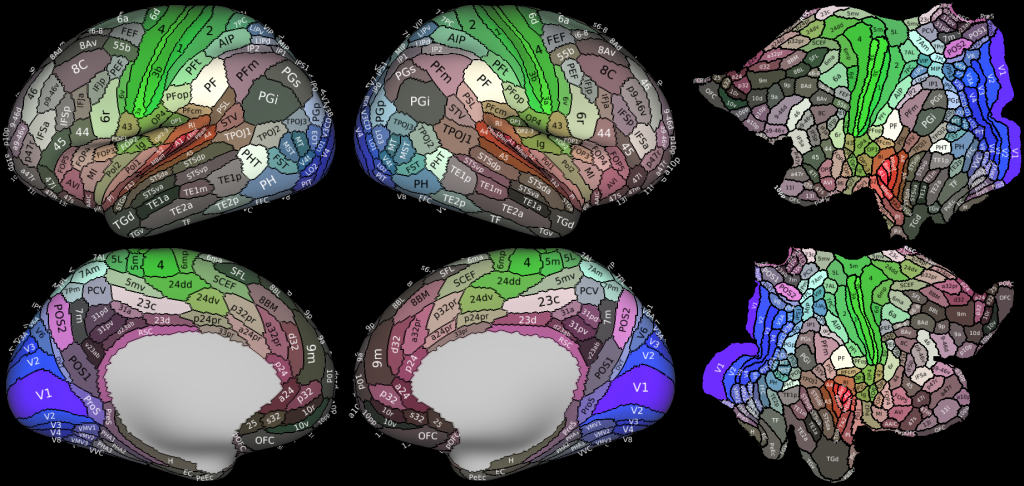
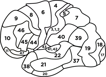
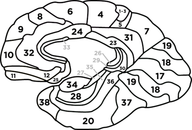
The 180 areas of the brain newly defined by the HCP2

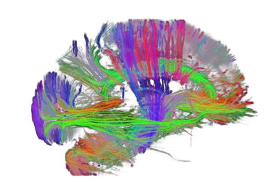
This parcellation also prompted groundbreaking findings on how these areas work together.

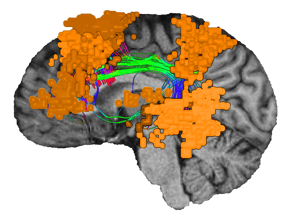
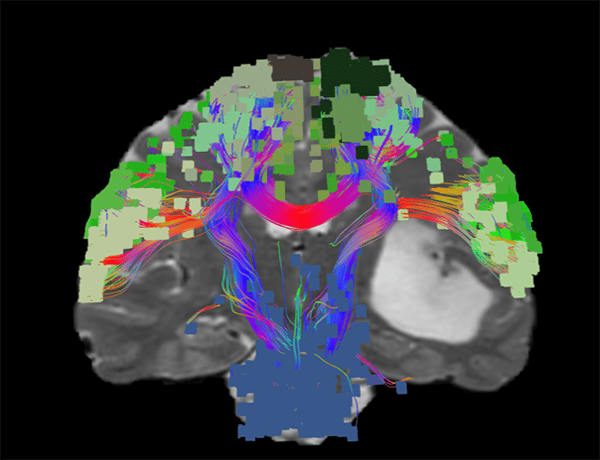
By studying the HCP parcellation, we now understand that cognition and function cannot be explained by individual regions. Instead, each functional area, or parcel, is a single node that participates in a larger brain network. These networks are responsible for important cognitive processes, like vision, movement, and internal thought. 

Imagine the brain as a modern day company

The company operates with multiple departments, such as product development, sales, marketing, HR, and others. On a typical day, each department has specific projects to complete, and every successful project contributes to the company’s overall operation. These departments are the main networks that operate in the human brain.

Now, take this analogy a step further: each department contains a team of employees working together on specific projects. In the sales department, one employee may search for new customers while another analyzes revenue data. While these two employees are in the same department, they perform different—although interrelated—tasks. The company’s employees are the individual nodes in the brain’s parcellation. While each node may have a primary function, its task processing is only a small part of a larger operation.

For too long, neuroscience focused on classifying individual nodes based on the tasks they performed, and this led to a misunderstanding of how the nodes work together. With a map of the brain and its connections, we’re now able to see not only how individual nodes function, but how they function together.

2021-02-11

Companies are about bringing together people of specific skill sets to accomplish massive and complex tasks - no one department or individual does it all. The brain is no different.

The Human Connectome Project's legacy

The HCP is far - very far - from the first effort to better understand brain connectivity and function having been established decades after many pivotal publications using techniques described in the following section. It did, however, mark a major turning point in consolidating a community and framework for computational brain mapping.

Its early success led to the launch of the Brain Research through Advancing Innovative Neurotechnologies (BRAIN) Initiative which has collectively led to the funding of hundreds of follow-up research projects amounting to over $1.5B USD in grant funding from five US Federal agencies: DARPA, NSF, IARPA, FDA, and NIH, as well as major industry, academic, and advocacy organizations.

Major breakthroughs have since led to fundamental shifts in our former understanding of the brain:

  • New models for brain disorders - the availability of this information and the techniques to acquire them allows for countless new dimensions and biomarkers to detect and treat brain disorders such as Alzheimer’s disease
  • The ability to study the brain as a network of nodes allows for applications of Graph Theory - similar to how the Google search engine optimizes its ability to find the most relevant information 
  • Brain network organisation - We can now relate brain network operation back to anatomy by determining what physical areas correspond to functional areas and their interconnections

In addition, organizations such as Omniscient  have been founded to bring these new findings and techniques into mainstream research and care through practical software that make such information easily accessible.

How to build a digital human brain map

Connectomics studies connectivity in the brain in two ways:

  1. Through its functional connections: which areas fire together
  2. Through its structural connections: which areas are wired together

With modern technology it is possible to obtain this for any person non-invasively from a single session with an MRI scanner. Combining these two forms of connectivity analysis with big data processing produces the single most comprehensive model of the human brain to date.

human brain map with connectomics

1. Mapping the functional

connectomics mapping the functional

When an area of the brain is engaged or in use, electrical activity tends to increase. We can model this with advanced MRI techniques like functional magnetic resonance imaging (fMRI). 

fMRI studies examine functional connectivity by measuring activity patterns through changes in cerebral blood flow. Basically, when a part of the brain is in use, it demands more oxygen and glucose that is carried by blood. Due to the magnetic properties of blood, these changes can be detected by MRI. We can observe patterns between functional areas in how they fire together and determine how they interrelate.  

2. Mapping the structural

connectomics mapping the structural

A 3D modeling technique known as tractography is then used to model data collected with another MRI-based technique called diffusion tensor imaging (DTI). Tractography models the path of water molecules as they move through nerve tracts in the brain’s white matter. When represented visually, the connective nerve tracts highlight how the brain’s different regions are physically “wired” together.

Collectively, these acquisition techniques provide an immense amount of data. Frankly, far too much for any individual to handle. Thus, from here, big data techniques including machine learning are used to combine functional and structural mapping to create an extremely comprehensive model of the human brain that is comprehensible.

The insights gleaned from such analysis have near limitless potential. 

Applying big data to the human brain

The amount of multi-dimensional data available in a digital brain parcellation is too complex for humans (even neuroscientists) to understand without big data processing techniques, such as machine learning and AI, and computing technologies, such as cloud computing.

With big data analytics, machine learning algorithms can conduct analysis on a parcellation’s dataset and identify patterns or make predictions about the human brain. This is similar to how algorithms can now detect fraudulent transactions or identify behavioral trends on platforms like Youtube.

These algorithms can also be trained to analyze subject-specific  data. Techniques such as those used in Omniscient software allow for the specific regions and variances of each individual to be mapped out and compared with, for example, normative data, and can also provide accurate localization of brain regions, no matter the differences in shape and structure.

How do you make sense of this?

There is a good chance that despite monumental shifts in our understanding of the brain resulting from Connectomics that you have not heard much about it. Much like the Human Genome Project and many other significant research projects preceding it, the HCP focused on discovery, not daily implementation. Creating detailed brain maps became possible, but not easy, and practical products usable by researchers and clinicians were not direct outputs of the project.

Instead, companies like Omniscient are further refining connectomic data with algorithms and technology to produce patient-specific datasets via dedicated, cloud-based software.

This means:

  • Standardizing and streamlining techniques born from over a decade of research to ensure consistent analyses across different sites
  • Partnering with MRI manufacturers to allow detailed mapping of brains from routine MRI scans
  • Harnessing cloud computing to processes vast volumes of data in reasonable time frames
  • Transforming this data into useful and actionable insights through intuitive product design that is rigorously tested

The resulting product is one where mapping an individual's brain is comparable to getting a blood test - both in how easy it is to do and how easy it is to interpret.

BN-language

This image shows the location of a subject's Language network determined by Omniscient software

Using Connectomics for neurological treatment

Dissecting the human brain into a parcellation that identifies functional areas and being able to study how these areas communicate is a profound advancement for neurological treatment.

Clinicians can gain critical information from a patient-specific parcellation that has never been available in the fields of neurosurgery and neurology. Since the brain is plastic and has the ability to change, we can use connectomic data to approach neurological illnesses and deficit as treatable conditions.

Using Connectomic data, neurosurgeons can with greater certainty understand what each incision in the brain will mean for a patient. Surgeons can adjust their approach to preserve the greatest amount of meaningful brain function.

connectomics for neurological treatment and neurosurgery

Analysis from Omniscient's Quicktome platform shows how a patient's motor networks organize around a brain tumor, and provide the neurosurgeon with important functional information prior to their first incision.

Rather than accepting Alzheimer’s disease or dementia as a normal part of aging. Better techniques for recognizing neurological degradation will lead to earlier diagnosis and the ability to slow the disease’s progression4. There is also far greater information available which will aid in the continued search for a cure.

Additionally, using a patient’s brain map to determine functional areas of deficit will allow physicians to provide patient-specific rehabilitation and treatment strategies. Such analysis may allow faster and more meaningful recovery of functional independence.

While these possibilities are incredible opportunities to treat neurological disease, advancements don’t have to be limited to neurological care. Connectomics can also be used for diagnosing and treating mental illness.

Applying Connectomics to mental illness

From a quantitative perspective, our understanding of mental illness remains extremely limited, often leading to non-definitive diagnoses and treatments. This is due to the tendencies of mental illnesses to present as disturbances of higher brain function that cannot be explained through physical examination alone.  That is, a typical MRI scan is unlikely to help diagnose mental illness as the biomarker is not structural like a tumor or stroke.

connectomics for neurosurgery and mental illness

On the left is a brain of a healthy individual. In the middle, this brain has a clear abnormality - a tumor. However, on the right - can you tell this patient has an anxiety disorder?

With almost a quarter of the world’s population set to face a mental illness at some stage of their life and hundreds of thousands of likely-preventable suicides occurring each year, the world’s mental health crisis is an incredibly pressing issue. In the case of mental illnesses, like PTSD, diagnosis typically requires identifying numerous co-occurring disorders and symptoms. In a similar manner, diseases like depression are actually multiple different disorders that exhibit similar symptoms5. Yet, they are often treated and diagnosed as one single disease.

To more effectively treat these complicated diseases, we must uncover the mechanisms that lead to mental illness, suicidal tendencies, and disorders. Using brain data analysis we can pinpoint these various symptoms and address them at their root cause of sickness in the brain.

Machine learning capabilities offer new opportunities to diagnose mental illness based on patterns of brain network function and connectivity. 

With fMRI scans, brain connectivity patterns can transform psychological disorders into a data problem. Machine learning algorithms can classify symptoms based on patterns of brain network dysfunction and make predictions about the symptom severity. Algorithms can also be trained to make comparisons between standard datasets and subject-specific data to diagnose patients with similarly-occurring symptoms. 

connectivity matrix brain

The image shown here is an Connectivity Matrix that compares a specific patient's functional connectivity to that of thousands of healthy datasets and finds patterns of anomaly - with red and blue regions indicating areas of abnormal behavior. This particular matrix was taken from the patient suffering an anxiety disorder in the last figure.

In terms of treatment, methods have also been found to correctly identify patients who are likely to respond positively to targeted neurostimulation therapies.

Using connectomic data, psychologists can begin pinpointing areas of disturbance in the brain, and far better understand what we currently call mental illness. As this research continues, these disorders are poised to become not only treatable but preventable.


Connectomics beyond medicine

While Connectomics offers a number of possibilities for neurological and neuropsychiatric care, its research potential does not have to stop there. The methods used to diagnose and treat deficits and disorders can provide valuable insight into our own minds. 

Generating and analyzing connectomic brain maps of healthy brains can help us understand our own intelligence, personality traits, and biases. This technology even presents the opportunity to enhance cognitive function and improve individual brain potential. Imagine identifying the brain networks responsible for certain skills (such as math and language), and being able to apply courses of non-invasive stimulation to improve them. 

This is not science fiction—studies have already shown promising results for brain enhancement.

Based on fMRI data, repetitive transcranial magnetic stimulation (rTMS)—targeted stimulation of key brain areas—has been shown to improve cognitive function for:

While these studies only scratch the surface of what is possible using non-invasive stimulation, they show that the possibilities are limitless.

The human brain is every individual’s most valuable asset, and little is more important than the future health and positive development of our own minds. With the connectomic data revolution, we are changing the future for every brain owner in the world.