---
title: The Sensorimotor Network
description: The sensorimotor network is the transducer, converting physical qualities like force, torque, pressure, or brightness into electrical signals as outputs.
image: https://www.o8t.com/hubfs/Sensorimotor-2.png
---

[![o8t_omniscient_Logo_RGB_Horizontal_MONO](https://www.o8t.com/hs-fs/hubfs/Omni_Logos/o8t_omniscient_Logo_RGB_Horizontal_MONO.png?width=174&height=48&name=o8t_omniscient_Logo_RGB_Horizontal_MONO.png) ![o8t_omniscient_Logo_RGB_Horizontal_REV_8000px](https://www.o8t.com/hs-fs/hubfs/o8t_omniscient_Logo_RGB_Horizontal_REV_8000px.png?width=174&height=39&name=o8t_omniscient_Logo_RGB_Horizontal_REV_8000px.png) ](https://www.o8t.com/)

[ Request a Demo ](https://www.o8t.com/book-a-demo)

[![o8t_omniscient_Logo_RGB_Horizontal_REV_8000px](https://www.o8t.com/hs-fs/hubfs/o8t_omniscient_Logo_RGB_Horizontal_REV_8000px.png?width=174&height=39&name=o8t_omniscient_Logo_RGB_Horizontal_REV_8000px.png) ](https://www.o8t.com/)

[ Request a Demo ](https://www.o8t.com/book-a-demo)

[Back to all blog posts ](https://www.o8t.com/blog)

[Brain Networks](https://www.o8t.com/blog/tag/brain-networks)

# The Sensorimotor Network

<https://www.o8t.com/blog/sensorimotor-network>

[Lewis Crawford](https://www.o8t.com/blog/author/lewis-crawford)

 2 June, 2022


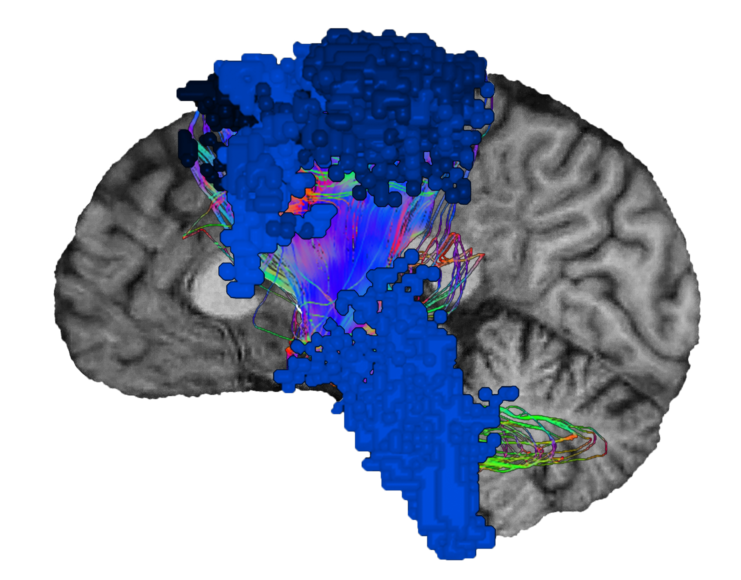
![BN-Sen](https://www.o8t.com/hs-fs/hubfs/BN-Sen.png?width=2467&name=BN-Sen.png)


#### Contents

- [What is the sensorimotor brain network? ](https://www.o8t.com/blog/sensorimotor-network#What-is-the-Sensorimotor-Network)
- [The role of the sensorimotor network ](https://www.o8t.com/blog/sensorimotor-network#The-role-Sensorimotor-Network)
- [The sensorimotor network’s role in bodily experience ](https://www.o8t.com/blog/sensorimotor-network#Sensorimotor-role-bodily-experience)
- [From discovery of the sensorimotor network to modern research ](https://www.o8t.com/blog/sensorimotor-network#discovery-to-research)
- [Where is the sensorimotor network located? ](https://www.o8t.com/blog/sensorimotor-network#Where-sensorimotor-network-located)
- [Close-knit interactions with other brain networks ](https://www.o8t.com/blog/sensorimotor-network#Close-knit-interactions-other-networks)
- [Irregularities in sensorimotor function ](https://www.o8t.com/blog/sensorimotor-network#Irregularities-in-sensorimotor-function)
- [A look to the future of sensorimotor study ](https://www.o8t.com/blog/sensorimotor-network#look-future-sensorimotor-study)
- [The main functional areas of the sensorimotor network ](https://www.o8t.com/blog/sensorimotor-network#Main-functional-areas-Sensorimotor)

## What is the sensorimotor brain network?

*The sensorimotor network is “the transducer.”*

As the brain's transducer, the sensorimotor network is responsible for sensing physical inputs, converting them to electrical signals that travel throughout the brain network, and then initiating a physical response

The sensorimotor network is responsible for:

- - Processing external physical stimuli
    - Feeling internal sensations
    - Evaluating the senses
    - Producing a motor response

## The role of the sensorimotor network

physicians pay special attention to the motor cortex, as well as the primary somatosensory cortex. Together with the secondary somatosensory cortex, the primary somatosensory cortex processes bodily sensations and sends signals to the motor cortex to execute reactions and appropriate motor responses.


## The sensorimotor network’s role in bodily experience

The sensorimotor network processes sensory inputs simultaneously to form a common experience from our senses.

Imagine sitting on a beach. You watch the tide roll in as the sunlight glistens on the water, and you hear the waves crash on the sand. Sitting in the sun also warms your body and makes your skin feel hot and sticky in the sand.

While the brain views these sensory perceptions as a single experience, different parts of your brain are responsible for analyzing and synthesizing these sensations.

The sensorimotor area oversees our response to external stimuli, like the feeling of sand, and the somatosensory cortex processes the experiences in and around the body, such as the warmth of the sun. Somatosensory reactions can be external, like temperature,1 pressure, and vibration,2 but they can also be internal proprioceptive sensations,3 like balance and coordination; or visceral senses,3 such as during indigestion or the pain experienced during irritable bowel syndrome.4

Since the sensorimotor network is one of the historically older brain networks, a swathe of study exists which investigates the varied aspects of physical sensation. Yet, the field of neuroscience is still making promising new discoveries as research focuses on the functional connectivity between the sensorimotor network and the brain’s other systems and subnetworks. To read more about functional brain networks, see [Brain Networks.](https://www.o8t.com/blog/brain-networks)

## From discovery of the sensorimotor network to modern research

Neurologists have long been interested in how the brain gives humans the ability to move and physically function. The earliest discovery of the sensorimotor network came about in 1870 when German neurologist, Eduard Hitzig, and anatomist, Gustav Fritsch, studied motor function in dogs.5They applied electrical stimulation to certain parts of the dogs’ cortex6 and identified regions where direct stimulation caused contralateral limb movements.5

Scottish neurologist, David Ferrier, also studied the effects of stimulation on the brain, but he expanded the scope of the stimulation to evoke movements of body parts from different regions of the cortex,7including the eyelids, face, mouth, hands, and feet.

**These studies and others paved the way for modern research, which has begun to evaluate the role the sensorimotor network plays in learning, skill acquisition, and memory.**

The primary motor cortex, somatosensory cortex, auditory and visual cortices have been shown to demonstrate plasticity during learning exercises with behavior validation.8 In addition to learned tasks during early developmental stages,10 learning exercises can include processes relating to attention, expectancy, learning strategy, and motivation in response to a learned signal stimulus.8

As the phenomenon of neuroplasticity has shown, physical experiences can result in changes to the brain's functional connectivity.10 Motor learning exercises, such as skilled finger movements,11 can lead to cortical reorganization in areas of the sensorimotor network responsible for motor learning and areas responsible for the perceptual changes that occur *during* motor learning.10

In addition to motor learning during waking moments, movements occuring during active (REM) sleep are thought to maintain and improve the sensorimotor system.12 Active sleep may also provide opportunity for cortical repair after sustaining an injury or during pathological states.12

Furthermore, repetitive transcranial magnetic stimulation (rTMS) has been shown to induce changes in sensory perception and motor performance in key areas of the sensorimotor network.13 Using rTMS to improve sensorimotor capabilities offers the possibility to modulate sensory perception and motor performance, and it poses new and exciting opportunities for neuroscience13 and treatment.

## Where is the sensorimotor network located?

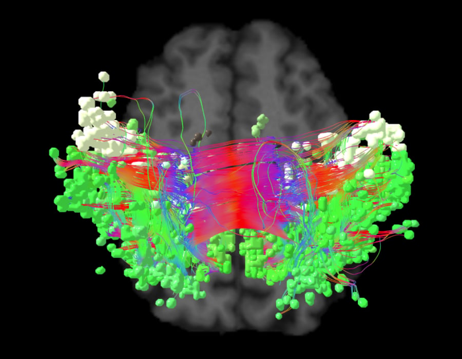
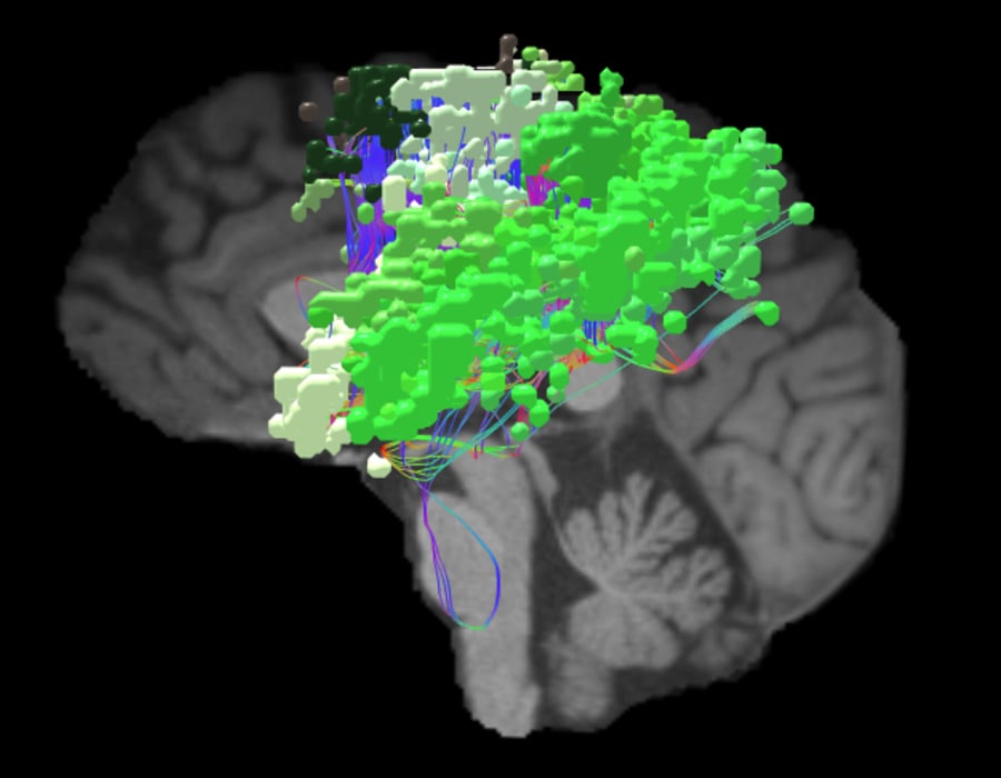
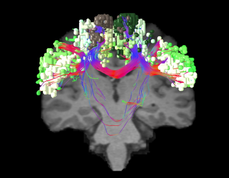
The sensorimotor network includes functional areas in the primary motor cortex, cingulate cortex, premotor cortex, and the supplementary motor area.14 It also includes the primary and sensory cortices in the parietal lobe.

While neuroscience has known the locations of motor planning regions since the late 19th century, using recent [connectomic data](https://www.o8t.com/blog/connectomics) from the Human Connectome Project15 has led to a more anatomically-specific understanding of the brain’s motor and somatic functional areas.


![SENSORIMOTOR_AXI](https://www.o8t.com/hubfs/Main%20Networks/3-POINT-VIEW%20IMAGES/SENSORIMOTOR_AXI.jpg)AXIAL VIEW

![SENSORIMOTOR_SAG](https://www.o8t.com/hubfs/Main%20Networks/3-POINT-VIEW%20IMAGES/SENSORIMOTOR_SAG.jpg) SAGITTAL VIEW

![SENSORIMOTOR_COR](https://www.o8t.com/hubfs/Main%20Networks/3-POINT-VIEW%20IMAGES/SENSORIMOTOR_COR.jpg) CORONAL VIEW

## Close-knit interactions with other brain networks

As the brain’s transducer, the sensorimotor network works closely with the other main [brain networks ](https://www.o8t.com/blog/brain-networks)to carry out key processes. It works alongside the:

- - Auditory subnetwork16 for hearing
    - [Visual system](https://www.o8t.com/blog/visual-system)17 for sight
    - [Limbic system](https://www.o8t.com/blog/limbic-system) for sense of smell18 and taste19
    - [Salience network](https://www.o8t.com/blog/salience-network) for behavior and reward processing20
    - [Central executive network](https://www.o8t.com/blog/central-executive-network) for task processing21
    - [Dorsal attention network](https://www.o8t.com/blog/dorsal-attention-network) for planning and controlling complex motor functions22,23
    - And the [default mode network](https://www.o8t.com/blog/default-mode-network) in cases of physical14 or mental illness24

Together, the individual nodes in these systems share functional connections that make up our senses and guide our actions. Yet, they can also lead to mental or physical illness when they become over- or underactive.

## Irregularities in sensorimotor function

Abnormalities in the sensorimotor network can cause sensory and movement disorders,25 as well as degenerative diseases, developmental delays, and mental health disorders.

Increased or decreased functional connectivity between brain networks is often associated with certain pathologies. Since the sensorimotor network is pervasive, it can be implicated in many different diseases and disorders.

The following are a few examples that may stem from irregularity in sensorimotor function:

Amyotrophic lateral sclerosis (ALS) patients with motor impairment, often exhibit reduced functional connectivity14 in the sensorimotor network. Network connectivity is also affected in patients with Huntington’s26 and Parkinson’s disease.27 These neurodegenerative diseases are some of the common manifestations of irregularities in sensorimotor function, yet the network is also implicated in less common disorders.

Since the [sensorimotor network](https://www.o8t.com/blog/sensorimotor-network) works closely with many subnetworks, irregularities in the network may also be present in problems of auditory perception.28

Likewise, decreased correspondence between the sensorimotor network and the visual cortex has been suggested to be a disturbance in perception-motor processing29 and may be a possible symptom of schizophrenia.

In mental health disorders, variable connectivity between the sensorimotor regions is present in individuals with autism spectrum disorder (ASD), and it has been shown to relate to their symptom severity.30,31 The relationship between the sensorimotor network and the [default mode network](https://www.o8t.com/blog/default-mode-network) may also play a factor in some mental health disorders. In the case of bipolar disorder,24 the default mode network can be overactive in states of depression until the sensorimotor network becomes overactive in manic states.24

Notably, increased sensorimotor connectivity potentially correlates with an increased non-responsiveness to antipsychotics.32 This could be a possible sign of treatment resistance for individuals with mental health disorders.

## A look to the future of sensorimotor study

Irregularities in the sensorimotor network can lead to many different physical and mental health disorders. Recent advances in neuroscience have uncovered cutting-edge opportunities for cortical reorganization that may improve abnormalities in the sensorimotor network.

Using rTMS, stimulating areas of the brain has the potential to promote behavioral changes, increased movement, and even sensory learning.33

In a 2008 study by Yoo et al.,13 rTMS was applied to the primary motor cortex to determine whether motor performance and sensory perception could be altered or improved during a sequential finger motor task. Using functional magnetic resonance imaging (fMRI), activation could be measured in specific areas of the brain, and areas of significant activation correlated with enhanced motor performance in subjects who received rTMS.

Other studies34 have found rTMS to evoke changes in stimulated brain areas for timeframes of at least 30 minutes.34 With additional research, these changes may offer longer-term improvements for sensory perceptions.

Beyond possible therapies, studying the sensorimotor network and our physical sensations can also help us understand an individual’s sense of self. A 2012 study35 presented participants with images of their own hand and the hand of a stranger. The representation of a person’s own hand appeared to be primarily confined to the left premotor cortex.35 This suggests that our understanding of our own body is an important part of the sensorimotor network.

The knowledge of our bodily self aids in differentiating ourselves from others. Continued study of the sensorimotor network may give new insight into a person's relationship with his or her own body, internal self, and the bodies of others.

Discover the key roles of other brain networks as they function alongside the sensorimotor network.

### The main functional areas in the sensorimotor network

Want to dive deeper?

[Full parcellation guide ](https://academy.o8t.com/parcellations/sensorimotor)

 1

###### Primary Sensorimotor Areas

The primary areas of the sensorimotor network are 1, 2, 3a, 3b, and 4. These nodes are responsible for processing tactile stimuli and sensory information from the skin. Area 4 is also implicated in fine motor movements and potentially plays a role in visual learning of motor-based skills early in life.

 2

###### Cingulate Cortex

Motor areas of the cingulate cortex include areas 24dd and 24dv which control complex motor planning and regulation for muscles through coordination with the supplementary motor area (SMA) and connections with the spinal cord. Area 24dd is responsible for muscles in the lower limb and trunk, whereas 24dv controls muscles in the upper limb and trunk. 

 3

###### Supplementary Motor Area (SMA)

The supplementary motor area (SMA) includes functional areas 6ma, 6mp, SCEF, and SFL. Areas 6ma and 6mp are activated when individuals receive visual instruction cues. Area SCEF is a higher-order oculomotor center implicated in goal-directed behavior. SFL is a hemispherical asymmetric area where in the left hemisphere the area is more active when listening to stories and responding to verbal cues, whereas in the right hemisphere, the area is activated in social interaction settings.

 4

###### Dorsal Premotor Cortex

Functional areas 6a and 6d in the dorsal premotor cortex associate informational cues with particular body movements. These may include solving math problems, interacting in social settings, and performing object feature comparison tasks, and they can be learned or based on visual or auditory sensation.

 5

###### Ventral Premotor Cortex

Area 6v in the ventral premotor cortex is a classic area related to the premotor cortex. It is involved in hand movements while manipulating objects, such as grasping or lifting. It also plays a role in learning actions or movements by first observing others performing a task. Area 6r in the ventral premotor cortex is also functionally related to Broca’s area, 55b, for language processing.

###### Share this article

- <https://www.facebook.com/sharer/sharer.php?u=https://www.o8t.com/blog/sensorimotor-network>
  
   Facebook
- <https://twitter.com/intent/tweet?url=https://www.o8t.com/blog/sensorimotor-network&text=The%20Sensorimotor%20Network>
  
   Twitter
- <http://www.linkedin.com/shareArticle?mini=true&url=https://www.o8t.com/blog/sensorimotor-network&title=The%20Sensorimotor%20Network&summary=The%20sensorimotor%20network%20is%20the%20transducer,%20converting%20physical%20qualities%20like%20force,%20torque,%20pressure,%20or%20brightness%20into%20electrical%20signals%20as%20outputs.>
  
   LinkedIn
- ###### Copy Link

###### related articles

## People who read this article also read

[Explore all articles ](https://www.o8t.com/blog)

<https://www.o8t.com/blog/the-trouble-with-calling-brain-regions-eloquent>

 June 2, 2022

[Brain Mapping](https://www.o8t.com/blog/topic/brain-mapping)

###### [The trouble with calling brain regions ‘eloquent’](https://www.o8t.com/blog/the-trouble-with-calling-brain-regions-eloquent)

<https://www.o8t.com/blog/the-auditory-system>

 June 2, 2022

[Brain Networks](https://www.o8t.com/blog/topic/brain-networks)

###### [The auditory system](https://www.o8t.com/blog/the-auditory-system)

<https://www.o8t.com/blog/ventral-attention-network>

 June 2, 2022

[Brain Networks](https://www.o8t.com/blog/topic/brain-networks)

###### [The Ventral Attention Network](https://www.o8t.com/blog/ventral-attention-network)

## Subscribe for important news and updates

You may unsubscribe from these communications at any time. For more information, check out our [privacy policy.](https://www.o8t.com/privacy-policy)

Reference List

##### Expand for full list of references

1. Donkelaar H.J., Broman J., van Domburg P. (2020) The Somatosensory System.*Clinical Neuroanatomy.*. Springer, Cham. [doi.org/10.1007/978-3-030-41878-6_4](https://link.springer.com/chapter/10.1007%2F978-3-030-41878-6_4#copyrightInformation)
2. Gleveckas-Martens N. Somatosensory system anatomy.*Medscape website. * [https://emedicine.medscape.com/article/1948621](https://emedicine.medscape.com/article/1948621.) Updated July 12, 2013. Accessed January 29, 2021. 
3. Feher J. 4.3 - Cutaneous sensory systems. In: Feher J, ed. Quantitative Human Physiology: An Introduction. *Cambridge, Academic Press; 2012:321-331.* [doi.org/10.1016/B978-0-12-382163-8.00035-9](https://www.sciencedirect.com/science/article/pii/B9780123821638000359?via%3Dihub) 
4. Icenhour A, Witt ST, Elsenbruch S, et al. Brain functional connectivity is associated with visceral sensitivity in women with Irritable Bowel Syndrome.*Neuroimage Clin. 2017. *[doi:10.1016/j.nicl.2017.06.001](https://www.researchgate.net/publication/317320728_Brain_functional_connectivity_is_associated_with_visceral_sensitivity_in_women_with_Irritable_Bowel_Syndrome)
5. Carlson C, Devinsky O. The excitable cerebral cortex Fritsch G, Hitzig E. Über die elektrische Erregbarkeit des Grosshirns.*Epilepsy Behav. 2009;15(2):123-30. *[http://citeseerx.ist.psu.edu/viewdoc/download?doi=10.1.1.704.6257&rep=rep1&type=pdf](http://citeseerx.ist.psu.edu/viewdoc/download?doi=10.1.1.704.6257&rep=rep1&type=pdf). Accessed January 29, 2021. 
6. Hagner M. The electrical excitability of the brain: toward the emergence of an experiment. *J Hist Neurosci.2012;21(3):237-49. * [doi: 10.1080/0964704X.2011.595634](https://pubmed.ncbi.nlm.nih.gov/22724486/) 
7. Taylor CSR, Gross CG. Twitches versus movements: A story of motor cortex. *Neuroscientist. 2003;9(5):332-342.* [doi.org/10.1177/1073858403257037](https://journals.sagepub.com/doi/10.1177/1073858403257037) 
8. Weinberger, N. 3.11 - Cortical plasticity in associative learning and memory. In: Byrne JH, ed. Learning and Memory: A Comprehensive Reference.*Cambridge, Mass: Academic Press; 2008;3:187-218.* [doi.org/10.1016/B978-012370509-9.00105-4](https://linkinghub.elsevier.com/retrieve/pii/B9780123705099001054) 
9. Del Rio‐Bermudez C, Blumberg MS. Active sleep promotes functional connectivity in developing sensorimotor networks.*Bioessays. 2018;40(4).* [doi.org/10.1002/bies.201700234](https://onlinelibrary.wiley.com/doi/abs/10.1002/bies.201700234) 
10. Vahdat S, Darainy M, Milner TE, Ostry DJ. Functionally specific changes in resting-state sensorimotor networks after motor learning.*J Neurosci. 2011;31(47):16907-16915.* [doi: 10.1523/JNEUROSCI.2737-11.2011.](https://www.jneurosci.org/content/31/47/16907) 
11. Andres FG, Mima T, Schulman AE, Dichgans J, Hallett M, Gerloff C. Functional coupling of human cortical sensorimotor areas during bimanual skill acquisition.* Brain. 1999;122(5):855-870. * [doi.org/10.1093/brain/122.5.855](https://academic.oup.com/brain/article/122/5/855/296636)
12. Blumberg MS. Developing sensorimotor systems in our sleep. *Curr Dir Psychol Sci. 2015;24(1):32-37.* [doi:10.1177/0963721414551362](https://journals.sagepub.com/doi/10.1177/0963721414551362)
13. BYoo W-K, You SH, Ko M-H, et al. High frequency rTMS modulation of the sensorimotor networks: Behavioral changes and fMRI correlates. *Neuroimage. 2008;39(4):1886-1895.* [doi.org/10.1016/j.neuroimage.2007.10.035](https://www.sciencedirect.com/science/article/abs/pii/S1053811907009937?via%3Dihub)
14. Chenji S, Jha S, Lee D, et al. Investigating default mode and sensorimotor network connectivity in amyotrophic lateral sclerosis.* PLoS One. 2016;11(6):e0157443.* [doi:10.1371/journal.pone.0157443](https://journals.plos.org/plosone/article?id=10.1371/journal.pone.0157443)
15. Human Connectome Project. Mapping the human brain connectivity. [www.humanconnectomeproject.org](http://www.humanconnectomeproject.org). Accessed October 16, 2020.  
16. Chang EF, Niziolek CA, Knight RT, Nagarajan SS, Houde JF. Human cortical feedback control of pitch.* Proc Natl Acad Sci U S A. 2013;110(7):2653-2658;* [doi:10.1073/pnas.1216827110](https://www.pnas.org/content/early/2013/01/22/1216827110) 
17. Comstock DC, Hove MJ, Balasubramaniam R. Sensorimotor synchronization with auditory and visual modalities: Behavioral and neural differences. *Front Comput Neurosci. 2018;12:53.* [doi:10.3389/fncom.2018.00053](https://www.frontiersin.org/articles/10.3389/fncom.2018.00053/full) 
18. Isaacson R. The Limbic System. Germany: Springer US; 2013. 
19. de Araujo IE, Simon SA. The gustatory cortex and multisensory integration. *Int J Obes. 2009;33:S34-S43.* [doi.org/10.1038/ijo.2009.70 ](https://www.nature.com/articles/ijo200970)
20. Olivo G, Wiemerslage L, Nilsson EK, et al. Resting-state brain and the FTO obesity risk allele: Default mode, sensorimotor, and salience network connectivity underlying different somatosensory integration and reward processing between genotypes. *Front Hum Neurosci. 2016;10:52.* [doi.org/10.3389/fnhum.2016.00052](https://www.frontiersin.org/articles/10.3389/fnhum.2016.00052/full)
21. Stevens MC, Kiehl KA, Pearlson GD, Calhoun VD. Functional neural networks underlying response inhibition in adolescents and adults. *Behav Brain Res. 2007;181(1):12-22.* [doi.org/10.1016/j.bbr.2007.03.023](https://www.sciencedirect.com/science/article/abs/pii/S0166432807001659?via%3Dihub)
22. Pi Y-L, Wu X-H, Wang F-J, et al. Motor skill learning induces brain network plasticity: A diffusion-tensor imaging study. *PLoS One. 2019;14(2):e0210015. *[doi.org/10.1371/journal.pone.0210015](https://journals.plos.org/plosone/article?id=10.1371/journal.pone.0210015)
23. Corbetta M, Shulman GL. Control of goal-directed and stimulus-driven attention in the brain. *Nat Rev Neurosci. 2002;3:201-215.* [doi.org/10.1038/nrn755](https://www.nature.com/articles/nrn755)
24. Doucet GE, Bassett DS, Yao N, Glahn DC, Frangou S. The role of intrinsic brain functional connectivity in vulnerability and resilience to bipolar disorder. *Am J Psychiatry. 2017;174(12):1214-1222.* [doi.org/10.1176/appi.ajp.2017.17010095](https://ajp.psychiatryonline.org/doi/10.1176/appi.ajp.2017.17010095)
25. Bologna M, Paparella G. Neurodegeneration and sensorimotor function. *Brain Sci. 2020;10(11):808.* [doi.org/10.3390/brainsci10110808](https://www.mdpi.com/2076-3425/10/11/808)
26. Dogan I, Eickhoff SB, Schulz JB. et al. Consistent neurodegeneration and its association with clinical progression in Huntington's disease: A coordinate-based meta-analysis. *Neurodegener Dis. 2013;12:23-35.* [doi.org/10.1159/000339528 ](https://www.karger.com/Article/Abstract/339528)
27. Sharman M, Valabregue R, Perlbarg V, et al. Parkinson's disease patients show reduced cortical‐subcortical sensorimotor connectivity. *Mov Disord. 2013;28:447-454.* [doi.org/10.1002/mds.25255 ](https://movementdisorders.onlinelibrary.wiley.com/doi/abs/10.1002/mds.25255)
28. Vanneste S, De Ridder D. The auditory and non-auditory brain areas involved in tinnitus. An emergent property of multiple parallel overlapping subnetworks. *Front Syst Neurosci. 2012;6:31.* [doi.org/10.3389/fnsys.2012.00031 ](https://www.frontiersin.org/articles/10.3389/fnsys.2012.00031/full)
29. Chen X, Duan M, Xie Q, et al. Functional disconnection between the visual cortex and the sensorimotor cortex suggests a potential mechanism for self-disorder in schizophrenia. *J Schizophr Res. 2015;166(1-3):151-157.* [doi.org/10.1016/j.schres.2015.06.014](https://www.sciencedirect.com/science/article/abs/pii/S0920996415003291?via%3Dihub)
30. Guo X, Duan X, Chen H, et al. Altered inter‐ and intrahemispheric functional connectivity dynamics in autistic children. *Hum Brain Mapp. 2020;41:419-428.* [doi.org/10.1002/hbm.24812](https://onlinelibrary.wiley.com/doi/full/10.1002/hbm.24812)
31. Harlalka V, Bapi RS, Vinod PK, Dipanjan R. Atypical flexibility in dynamic functional connectivity quantifies the severity in autism spectrum disorder. *Front Hum Neurosci. 2019;13:6.* [doi.org/10.3389/fnhum.2019.00006](https://www.frontiersin.org/articles/10.3389/fnhum.2019.00006/full)
32. McNabb CB, Sundram F, Soosay I, Kydd RR, Russell BR. Increased sensorimotor network connectivity associated with clozapine eligibility in people with schizophrenia. *Psychiatry Res Neuroimaging. 2018;275:36-42.* [doi.org/10.1016/j.pscychresns.2018.02.008](https://www.sciencedirect.com/science/article/abs/pii/S092549271730286X?via%3Dihub)
33. Heba S, Lenz M, Kalisch T, et al. Regionally specific regulation of sensorimotor network connectivity following tactile improvement. *Neural Plast. 2017;2017:1-11.* [doi.org/10.1155/2017/5270532](https://www.hindawi.com/journals/np/2017/5270532/)
34. Ragert P, Franzkowiak S, Schwenkrei P, Tegenthoff M, Dinse HR. Improvement of tactile perception and enhancement of cortical excitability through intermittent theta burst rTMS over human primary somatosensory cortex. *Exp Brain Res. 2008;184:1-11.* [doi.org/10.1007/s00221-007-1073-2 ](https://link.springer.com/article/10.1007/s00221-007-1073-2)
35. Ferri F, Frassinetti F, Ardizzi M, Costantini M, Gallese V. A sensorimotor network for the bodily self. *J Cogn Neurosci. 2012;24(7):1584-1595.* [doi.org/10.1162/jocn_a_00230 ](https://www.mitpressjournals.org/doi/abs/10.1162/jocn_a_00230)

[![Bitmap Copy](https://www.o8t.com/hubfs/Salted%20Stone/images/Bitmap%20Copy.svg) ](https://www.o8t.com)

![Badges_4](https://www.o8t.com/hs-fs/hubfs/Badges_4.webp?width=200&name=Badges_4.webp)![soc-2-1](https://www.o8t.com/hs-fs/hubfs/soc-2-1.webp?width=67&height=86&name=soc-2-1.webp)       
© o8t 2026  |  [Privacy Policy](https://www.o8t.com/privacy-policy)

Follow us on:

<https://www.linkedin.com/company/omniscientneurotechnology> <https://twitter.com/o8tneuro> <https://www.youtube.com/channel/UCFth5-xQd5VSm1dD7sNcZ3Q/featured>

```json
{
  "@context" : "https://schema.org",
  "@type" : "BlogPosting",
  "author" : {
    "@type" : "Person",
    "name" : "Lewis Crawford",
    "url" : "https://www.o8t.com/blog/author/lewis-crawford"
  },
  "dateModified" : "2022-10-13T05:35:39.943Z",
  "datePublished" : "2022-06-02T22:37:00.000Z",
  "headline" : "The Sensorimotor Network",
  "image" : [ "https://www.o8t.com/hubfs/Sensorimotor-2.png" ],
  "mainEntityOfPage" : {
    "@id" : "https://www.o8t.com/blog/sensorimotor-network",
    "@type" : "WebPage"
  },
  "publisher" : {
    "@type" : "Organization",
    "logo" : {
      "@type" : "ImageObject",
      "url" : "https://www.o8t.com/hubfs/Omni_Logos/o8t_omniscient_Logo_RGB_Horizontal_MONO.png"
    },
    "name" : "Omniscient Neurotechnology"
  }
}
```