Hyper-personalized brain care
In a single scan, Quicktome brings brain networks to light – equipping you with patient-specific, actionable insight.
The most powerful scan you’ve never seen
Quicktome is intended for brain health physicians who demand more knowledge of their patient’s brain to drive better care.
A powerful cloud-based solution, Quicktome delivers fast and intuitive insights into a patient’s brain networks. We harness rich data from a single, non-invasive MRI scan, process them automatically, and deliver them in an intuitive web-based app.
Quicktome enables:
- https://www.ncbi.nlm.nih.gov/pmc/articles/PMC9029431
- https://academic.oup.com/noa/article/4/1/vdac142/6705399


3. https://pubmed.ncbi.nlm.nih.gov/30639605/
4. 100% of surveyed neurosurgeons in the Quicktome Post Market Clinical Survey agreed that Quicktome helps them communicate with their patients more effectively.
FEATURES
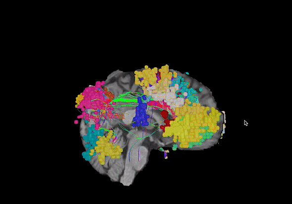
Brain maps made for every case
.gif?width=410&height=281&name=Product%20page%20SC%26FC%20(1).gif)
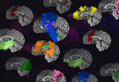
Structural and Functional Brain Mapping
Switch between insights generated by white matter analyses (tractography) or whole-brain functional mapping (rs-fMRI)*.
*Feature may not yet be available in your region.
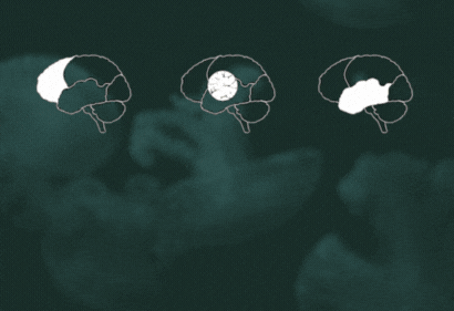
Network Selection
We distill decades of research to help you easily select and interrogate brain networks, tract bundles, and individual brain parcels.
Workflow Presets
Interactive Connectome Guide
Compatible exports
All maps can be exported to third-party DICOM (Digital Imaging and Communications in Medicine) compatible devices.
Personalized, because it matters
Tap the yellow dots to find out more
Automatic edema correction
Mitigates processing noise related to cerebral edema allowing brain networks to be mapped in critical regions, such as around a brain tumor prior to surgery.
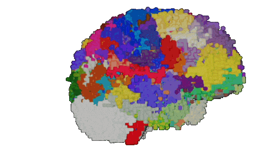
360 functional areas1 mapped with SCA technology2
Omniscient’s Structural Connectivity Atlas (SCA) uses machine learning and tractographic techniques to create highly specific and personalized maps of the human brain.
Industry-leading noise correction
Raw MRI scans undergo a series of steps that correct noise, including gradient distortion correction and subject motion correction.
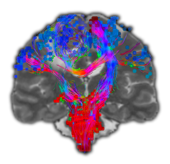
Next-generation tractography via CSD3
Constrained Spherical Deconvolution (CSD) is superior to traditional Diffusion Tensor Imaging, especially in areas of crossing and branching white matter, allowing clinicians to be more precise in preserving and targeting functional areas.
Automatic edema correction
Mitigates processing noise related to cerebral edema allowing brain networks to be mapped in critical regions, such as around a brain tumor prior to surgery.
360 functional areas1 mapped with SCA technology2
Omniscient’s Structural Connectivity Atlas (SCA) uses machine learning and tractographic techniques to create highly specific and personalized maps of the human brain.
References
Industry-leading noise correction
Raw MRI scans undergo a series of steps that correct noise, including gradient distortion correction and subject motion correction.
Next-generation tractography via CSD3
Constrained Spherical Deconvolution (CSD) is superior to traditional Diffusion Tensor Imaging, especially in areas of crossing and branching white matter, allowing clinicians to be more precise in preserving and targeting functional areas.
References
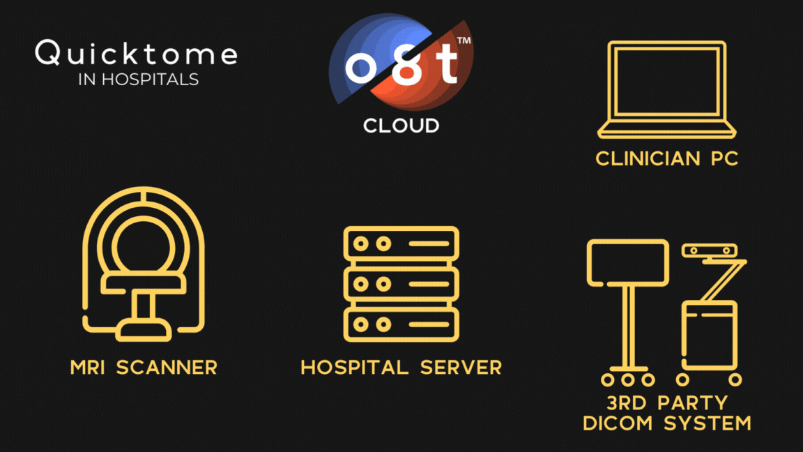
INTEGRATIONS
Bringing the most advanced brain imaging to you
APPLICATIONS
Enabling new possibilities in brain health


Neurosurgeons
Quicktome backs each incision with knowledge of a patient’s brain networks, preserving functional independence and maximizing onco-functional balance.
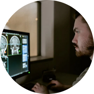
Neuroradiologists
With automated resting-state fMRI insights for whole brain functional analysis and personalized connectome mapping, Quicktome enables a Hybrid Intelligence approach to neuroradiology, combining AI with human expertise.

Hospital administration
Quicktome supercharges brain health services by delivering crucial brain network insights – all without new capital equipment or maintenance fees.
Neurosurgeons
Quicktome backs each incision with knowledge of a patient’s brain networks, preserving functional independence and maximizing onco-functional balance.
Neuroradiologists
With automated resting-state fMRI insights for whole brain functional analysis and personalized connectome mapping, Quicktome enables a Hybrid Intelligence approach to neuroradiology, combining AI with human expertise.
Hospital administration
Quicktome supercharges brain health services by delivering crucial brain network insights – all without new capital equipment or maintenance fees.
Interested in more information?
Fill out this form and a member of the Omniscient team will get in touch with you shortly.


© o8t 2026 | Privacy Policy