Key Highlights
  • To achieve an optimal onco-functional balance in neurosurgery, a more comprehensive appreciation of brain anatomy and function is paramount. 
  • Brain networks underpin a range of human functions, many of which overlap with deficits following neurosurgery.
  • Personalized brain mapping visualizes brain networks and associated tract bundles in individual patients, providing a practical solution for intraoperative navigation and formulating pre-surgical approaches.

Introduction

The absolute goal of intracerebral neurosurgery, especially when tumors are involved, is to provide optimal treatment whilst preserving patient quality-of-life1,2. How aggressive a surgeon can be in removing tumor-affected tissue without compromising regular functions like movement, cognition, or emotion, is often referred to as “onco-functional balance”3. In order to maximize onco-functional balance, an understanding of patient’s unique brain anatomy prior to surgery is required - a requirement met by personalized brain mapping4.  

It is widely accepted that the greatest chance of extending life in tumor patients comes from supramaximal resection3,5. However, these approaches have also, albeit rarely, shown to lead to devastating and unpredicted functional consequences for patients after surgery6–9. These consequences can largely be explained by incidental or necessary damage to brain networks: connected groups of functional brain regions. Personalized brain mapping is a technique capable of locating these brain networks pre-surgically, and further, assess their composition post surgery to better understand how individual patients recover after surgery10–12.   

Conceptually, brain networks originated in academia over 20 years ago, with the Default Mode Network (DMN) - the network responsible for at-rest consciousness - the first to be discovered13. Through ongoing research and academic insight, we now understand that unique brain networks hold critical roles in a range of executive functions including movement (sensorimotor network), cognition (central executive network), and vision (visual system), amongst others. Figure 1 shows each of the 7 major brain networks displayed in Quicktome software. Despite their lengthy history in academia, understanding of brain networks rarely reaches the surgical suite - primarily due to the inability to reliably define their location in an individual patient. Personalized brain mapping removes this barrier, for the first time bringing the forefront of academic understanding into a clinical relevant landscape. 

What makes personalized brain mapping “personalized”?

Modern understanding of the brain anatomy emerged from the 2016 Human Connectome Project - a multisite, multidisciplinary project which produced an atlas of the brain comprised of 180 regions per hemisphere. Their nomenclature was later used by Baker, Briggs, and Sughrue to define each of the brain’s major networks in their “Connectomic Atlas of the Human Cerebrum”. 14,15 This atlas was both granular enough to discern the discrete function of each brain region, yet simple enough to be discussed and applied between institutions - enabling a common language for discussing brain anatomy. 

Personalized brain mapping works by visualizing where each of the functional areas described within this atlas lie in an individual, rather than assuming that a single archetypal atlas is applicable in every case. By employing a machine-learning approach to process brain data, personalized brain mapping generates a unique anatomical map of each functional brain area and the tracts which connect them, regardless of variations in gyral folding patterns, intracranial volume, or structural pathology. An in-depth description of the method for conducting personalized brain mapping has been described in the work of Doyen et al. and their Structural Connectivity Atlas (SCA),16 however, can be summarized as:

  1. Generating Connectivity Profiles: Train a machine-learning algorithm using Diffusion Weighted Imaging (DWI) data to identify how white matter tracts connect each atlas region in a healthy individual (structural connectivity).

  2. Collecting Patient Data: Collect and process patient DWI data, tracing out the location, distribution, and termination points of their individual white matter tracts.

  3. Model application: Enter patient DWI data into the algorithm trained in (1.), which recreates the atlas in patient space, marking out region locations based on how a patient’s white matter tracts are distributed and connected.

Figure 3 transparent-1Figure 2. Schematic description of a personalized brain mapping method (Structural Connectivity Atlas / SCA). By generating a machine learning algorithm using healthy brain data, personalized brain mapping seeks to create patient-specific representations of complex brain atlases. This method utilizes a modified version of the Human Connectome Project (HCP) atlas, and re-parcellates this atlas on a patient's brain, as informed by underlying structural connectivity.

How personalized brain mapping informs neurosurgical decision making

Personalized brain mapping incorporates a methodology which innately promotes its use in surgical settings. First and foremost, the ability to visualize and freely segment named brain networks and associated tract bundles is of great importance in planning surgical trajectories. Additionally however, personalized brain mapping brings into the hands of a surgeon: 1) a greater clarity of brain network involvement in surgical pathology pre-operatively, 2) the ability to structurally inform maximal safe resection intraoperatively, and most importantly, 3) enhanced predictions of potential postoperative consequences, generating a complete informed consent for patients. 


Brain network involvement 

Pre-operative assessment of a patient’s brain networks gives surgeons a thorough understanding of how pathology has already impacted functional brain networks. Damage to these networks can cause deficits in the functions in which they underpin or manifest entirely new cognitive or neuropsychiatric disturbances. 

Taking for example the three main cognitive brain networks – the Central Executive (CEN), Default Mode (DMN), and Salience Network (SN). The CEN drives goal-directed behavior, and conversely, the DMN activates during passive states of mind and at rest17-19. The balance of switching between these two networks during daily life is mediated by the SN20. Unsurprisingly, damage to or disconnection of the tracts between these major networks can cause impairments to higher-order cognitive abilities and cause symptoms associated with schizophrenia, depression, and anxiety21-23. The use of personalized brain mapping preoperatively informs neurosurgeons as to the precise location and involvement of these networks, among others, in their patient’s pathology, allowing them to plan accordingly to avoid and navigate these networks during surgery24–26


Maximal safe resection 

The precise anatomical visualizations produced by personalized brain mapping empower neurosurgeons seeking a safe maximal resection. As they are informed by tracts connecting functional gray matter areas, not only can a surgeon use personalized brain mapping to see where a tumor site is entangled with functioning brain tissue, but also if adjacent gray matter has already become compromised by a tumor’s presence prior to surgery. This information not only informs surgeons which still-functioning tracts and networks to avoid in their approach, but also which already compromised tissue can be safely resected without leading to worse post-operative symptoms27-29


Better patient consent

Since the 1940’s, consent laws have been in place to ensure that patients are made fully aware of the potential risk-factors of intracranial procedures30. By displaying the location of brain networks and their connections relative to structural damage, personalized brain mapping enables a neurosurgeon to directly demonstrate to their patient where and why performing the elected surgical strategy could result in postsurgical deficits1,4. Instances where a patient has awoken from surgery with an unexplained deficit have shown to result from components of core brain networks being damaged in the surgical approach31-35.  What is of paramount importance in cases such as these is that patients are made aware of how their deficits relate to changes in their brain’s structure and function - which is made possible by personalized brain mapping36,37.


Picture 4

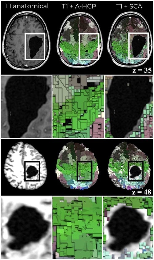
Figure 3. Personalized brain mapping resolving functional areas around structural pathology. Left column: a T1-weighted image showing the location and extent of structural damage. Middle column: a typical brain atlas overlayed on the T1 image, demonstrating an inability to account for structural damage. Right column: The Structural Connectivity Atlas (SCA) personalized brain mapping technique, showing that brain regions are mapped in accordance and surrounding structural damage.

Conclusion

Omniscient Neurotechnology’s Quicktome software enables a personalized brain mapping solution which is accessible by collecting basic MRI scanning sequences. By loading an anatomical and Diffusion Weighted Imaging sequence into Quicktome, neurosurgeons can view and segment out individual brain networks and tracts associated with their patients structural pathology or symptom profile, or utilize the inbuilt pre-selection tools for damage isolated to any major lobe of the brain. The personalized brain maps generated by Quicktome are easily integrated with modern PACS systems, providing neurosurgeons with actionable insights prior to, during, and following their chosen surgical trajectory.

Download the whitepaper on Network-Based Neurosurgery

Whitepaper-cover_400px

Personalizing Neurosurgery: How Understanding Brain Networks Aids the Surgical Approach

Read more about maximizing onco-functional balance in neurosurgery with personalized brain mapping.

References
Expand to see full list of references
  1. Dadario NB, Brahimaj B, Yeung J, Sughrue ME. Reducing the Cognitive Footprint of Brain Tumor Surgery. Frontiers in Neurology. 2021;0:1342. doi:10.3389/FNEUR.2021.711646
  2. Almeida JP, Chaichana KL, Rincon-Torroella J, Quinones-Hinojosa A. The Value of Extent of Resection of Glioblastomas: Clinical Evidence and Current Approach. Current Neurology and Neuroscience Reports. 2015;15(2):1-13. doi:10.1007/S11910-014-0517-X/FIGURES/4
  3. Molinaro AM, Hervey-Jumper S, Morshed RA, et al. Association of Maximal Extent of Resection of Contrast-Enhanced and Non-Contrast-Enhanced Tumor With Survival Within Molecular Subgroups of Patients With Newly Diagnosed Glioblastoma. JAMA Oncol. 2020;6(4):495-503. doi:10.1001/JAMAONCOL.2019.6143
  4. Yeung JT, Taylor HM, Nicholas PJ, et al. Using Quicktome for intracerebral surgery: early retrospective study and proof of concept. World Neurosurgery. Published online August 3, 2021. doi:10.1016/j.wneu.2021.07.127
  5. Li YM, Suki D, Hess K, Sawaya R. The influence of maximum safe resection of glioblastoma on survival in 1229 patients: Can we do better than gross-total resection? Journal of Neurosurgery. 2016;124(4):977-988. doi:10.3171/2015.5.JNS142087
  6. Herbet G, Moritz-Gasser S. Beyond Language: Mapping Cognition and Emotion. Neurosurgery Clinics of North America. 2019;30(1):75-83. doi:10.1016/J.NEC.2018.08.004
  7. Drewes C, Sagberg LM, Jakola AS, Solheim O. Perioperative and Postoperative Quality of Life in Patients with Glioma–A Longitudinal Cohort Study. World Neurosurgery. 2018;117:e465-e474. doi:10.1016/J.WNEU.2018.06.052
  8. Rijnen SJM, Kaya G, Gehring K, et al. Cognitive functioning in patients with low-grade glioma: effects of hemispheric tumor location and surgical procedure. Journal of Neurosurgery. 2019;133(6):1671-1682. doi:10.3171/2019.8.JNS191667
  9. Starnoni D, Berthiller J, Idriceanu TM, et al. Returning to work after multimodal treatment in glioblastoma patients. Neurosurgical Focus. 2018;44(6):E17. doi:10.3171/2018.3.FOCUS1819
  10. Noll KR, Sullaway CM, Wefel JS. Depressive symptoms and executive function in relation to survival in patients with glioblastoma. Journal of Neuro-Oncology. 2019;142(1):183-191. doi:10.1007/S11060-018-03081-Z/TABLES/4
  11. Vassal M, Charroud C, Deverdun J, et al. Recovery of functional connectivity of the sensorimotor network after surgery for diffuse low-grade gliomas involving the supplementary motor area. Journal of Neurosurgery. 2017;126(4):1181-1190. doi:10.3171/2016.4.JNS152484
  12. Carbo EWS, Hillebrand A, van Dellen E, et al. Dynamic hub load predicts cognitive decline after resective neurosurgery. Scientific Reports 2017 7:1. 2017;7(1):1-10. doi:10.1038/srep42117
  13. Raichle ME, Macleod AM, Snyder AZ, Powers WJ, Gusnard DA, Shulman GL. A default mode of brain function. National Academy of Sciences. Published online 1996. Accessed April 18, 2022. www.pnas.org
  14. Baker CM, Burks JD, Briggs RG, et al. A Connectomic Atlas of the Human Cerebrum—Chapter 1: Introduction, Methods, and Significance. Operative Neurosurgery. 2018;15(Suppl 1):S1. doi:10.1093/ONS/OPY253
  15. Glasser MF, Coalson TS, Robinson EC, et al. A multi-modal parcellation of human cerebral cortex. Nature 2016 536:7615. 2016;536(7615):171-178. doi:10.1038/nature18933
  16. Doyen S, Nicholas P, Poologaindran A, et al. Connectivity-based parcellation of normal and anatomically distorted human cerebral cortex. Hum Brain Mapp. Published online 2021. doi:10.1002/HBM.25728
  17. Menon V. The Triple Network Model, Insight, and Large-Scale Brain Organization in Autism. Biological Psychiatry. 2018;84(4):236-238. doi:10.1016/J.BIOPSYCH.2018.06.012
  18. Sandhu Z, Tanglay O, Young IM, et al. Parcellation-based anatomic modeling of the default mode network. Brain and Behavior. 2021;11(2):e01976. doi:10.1002/BRB3.1976
  19. Cao W, Cao X, Hou C, et al. Effects of cognitive training on resting-state functional connectivity of default mode, salience, and central executive networks. Frontiers in Aging Neuroscience. 2016;8(APR):70. doi:10.3389/FNAGI.2016.00070/BIBTEX
  20. Goulden N, Khusnulina A, Davis NJ, et al. The salience network is responsible for switching between the default mode network and the central executive network: Replication from DCM. Neuroimage. 2014;99:180-190. doi:10.1016/J.NEUROIMAGE.2014.05.052
  21. Chen H, Li Y, Liu Q, et al. Abnormal interactions of the salience network, central executive network, and default-mode network in patients with different cognitive impairment loads caused by leukoaraiosis. Frontiers in Neural Circuits. 2019;13:42. doi:10.3389/FNCIR.2019.00042/BIBTEX
  22. Mohan A, Roberto AJ, Mohan A, et al. Focus: The Aging Brain: The Significance of the Default Mode Network (DMN) in Neurological and Neuropsychiatric Disorders: A Review. The Yale Journal of Biology and Medicine. 2016;89(1):49. Accessed April 3, 2022. /pmc/articles/PMC4797836/
  23. Pannekoek JN, van der Werff SJ, Meens PH, et al. Aberrant resting-state functional connectivity in limbic and salience networks in treatment--naïve clinically depressed adolescents. J Child Psychol Psychiatry. 2014;55(12):1317-1327. doi:10.1111/jcpp.12266
  24. Beevers CG. Cognitive vulnerability to depression: A dual process model. Clinical Psychology Review. 2005;25(7):975-1002. doi:10.1016/J.CPR.2005.03.003
  25. Ren H, Zhu J, Su X, et al. Application of Structural and Functional Connectome Mismatch for Classification and Individualized Therapy in Alzheimer Disease. Frontiers in Public Health. 2020;8:720. doi:10.3389/FPUBH.2020.584430/BIBTEX
  26. Zhou L, Pu W, Wang J, et al. Inefficient DMN Suppression in Schizophrenia Patients with Impaired Cognitive Function but not Patients with Preserved Cognitive Function. Scientific Reports 2016 6:1. 2016;6(1):1-10. doi:10.1038/srep21657
  27. Dubey A, Kataria R, Sinha VD. Role of Diffusion Tensor Imaging in Brain Tumor Surgery. Asian J Neurosurg. 2018;13(2):302-306. doi:10.4103/ajns.AJNS_226_16
  28. Sparacia G, Parla G, Lo Re V, et al. Resting-State Functional Connectome in Patients with Brain Tumors Before and After Surgical Resection. World Neurosurg. 2020;141:e182-e194. doi:10.1016/j.wneu.2020.05.054
  29. Morales H. Current and Future Challenges of Functional MRI and Diffusion Tractography in the Surgical Setting: From Eloquent Brain Mapping to Neural Plasticity. Semin Ultrasound CT MR. 2021;42(5):474-489. doi:10.1053/j.sult.2021.07.005
  30. Ammar A, Bernstein M. Neurosurgical Ethics in Practice: Value-based Medicine 123.
  31. Martin HC, Sethi J, Lang D, Neil-Dwyer G, Lutman ME, Yardley L. Patient-assessed outcomes after excision of acoustic neuroma: postoperative symptoms and quality of life. J Neurosurg. 2001;94(2):211-216. doi:10.3171/jns.2001.94.2.0211
  32. Lopez Ramos C, Brandel MG, Rennert RC, et al. Clinical Risk Factors and Postoperative Complications Associated with Unplanned Hospital Readmissions After Cranial Neurosurgery. World Neurosurg. 2018;119:e294-e300. doi:10.1016/j.wneu.2018.07.136
  33. Daabiss M. American Society of Anaesthesiologists physical status classification. Indian J Anaesth. 2011;55(2):111-115. doi:10.4103/0019-5049.79879
  34. Campanella F, Fabbro F, Ius T, Shallice T, Skrap M. Acute effects of surgery on emotion and personality of brain tumor patients: surgery impact, histological aspects, and recovery. Neuro Oncol. 2015;17(8):1121-1131. doi:10.1093/neuonc/nov065
  35. Campanella F, Shallice T, Ius T, Fabbro F, Skrap M. Impact of brain tumour location on emotion and personality: a voxel-based lesion-symptom mapping study on mentalization processes. Brain. 2014;137(Pt 9):2532-2545. doi:10.1093/brain/awu183
  36. Briggs RG, Allan PG, Poologaindran A, et al. The Frontal Aslant Tract and Supplementary Motor Area Syndrome: Moving towards a Connectomic Initiation Axis. Cancers 2021, Vol 13, Page 1116. 2021;13(5):1116. doi:10.3390/CANCERS13051116
  37. Baker CM, Burks JD, Briggs RG, et al. The crossed frontal aslant tract: A possible pathway involved in the recovery of supplementary motor area syndrome. Brain and Behavior. 2018;8(3):e00926. doi:10.1002/BRB3.926