Key Highlights
  • A 34-year-old male presented with seizures. A subsequent MRI revealed a hyperintense infiltrative lesion compromising the left superior frontal gyrus.
  • Quicktome analysis revealed involvement of the frontal aslant tract (FAT) in the supplementary motor area (SMA), and the corticospinal tract (CST).
  • Awake mapping was deemed not possible due to the patient's anesthesiologic contraindications.
  • Following craniotomy, the posterior half of the superior frontal gyrus and medial precentral gyrus were exposed.
  • Intra-operative MRI confirmed the adequate resection, preserving the CST.

Patient history

A 34-year-old male patient presenting a first seizure underwent investigation. MRI showed a FLAIR hyperintense infiltrative lesion compromising left superior frontal gyrus. There was minimal T1 contrast enhancement. Spectroscopy was compatible with a glial tumor, with ‘hot spots’ in the perfusion suggesting a high-grade lesion, with possibility of oligodendroglial lineage. He had a normal neurological and neuropsychological evaluation.

Figure 1

Brain networks as seen in Quicktome: Sagittal view revealing the tumor posterior limit, with the supplementary motor area (SMA) of the corticospinal tract (CST).

Brain network involvement

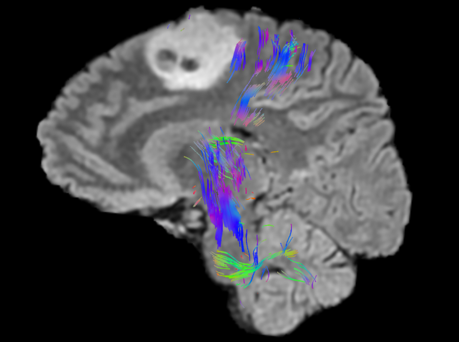
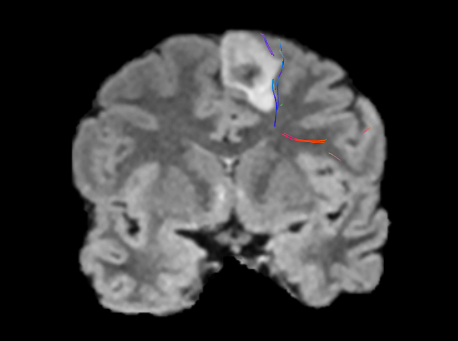
Quicktome was used to evaluate the relationship of the tumor with the surrounding areas, particularly the frontal aslant tract (FAT) in the supplementary motor area (SMA), and the corticospinal tract (CST).

These areas were in close proximity to the tumor and so extra care was needed to avoid damage to the FAT, which can result in speech disorders, as well as impairment of executive functions, visual–motor activities, orofacial movements, inhibitory control, working memory, social community tasks, attention, and music processing. Damage to the CST can lead to an irreversible contralateral motor deficit, and surgical manipulation of the SMA tends to cause transient deficits, recoverable within a few weeks. It is a well-known complication of medial frontal lobe surgery called SMA syndrome.

Performing surgery awake was deemed not possible due to the patient's anesthesiologic contraindications.

Figure 2

Coronal view in Quicktome showing the lateral boundary of the tumor with the frontal aslant tract.

Surgical decisions and outcomes

After craniotomy, the posterior half of the superior frontal gyrus and medial precentral gyrus were exposed. 5-ALA fluorescence was positive in small spots of the tumor. Intra-operative MRI confirmed the adequate resection, preserving the CST. In the immediate postoperative period, the patient presented hemiparesis in the right side of the body, with progressive and complete recovery after 10 days. There was no speech deficit. Final diagnosis confirmed astrocytoma WHO grade 2 IDH-wildtype, demanding aggressive adjuvant treatment with irradiation and temozolomide.

Download the case study

CAS0006 Image

Recurrent glioblastoma and restoring speech function

Download the case study in PDF format.

SEE THE QUICKTOME DIFFERENCE

Upload a free test case, or ask us how Quicktome could work for your site.