Key Highlights
  • Quicktome was used to analyse both hemispheres, comparing disrupted fibers to normal fibers
  • Quicktome was paired with intraoperative asleep motor mapping to confirm the fibers that needed preservation
  • Motor function improved post-operatively, due to the tumor resection which subsequently relieved pressure on the fibers

Patient history

A young lady with complaints of acute left leg weakness arrived at hospital with zero strength in her left leg. Imaging revealed an approx. 3cm mass in her primary motor strip.

Brain network involvement

Quicktome brain mapping software was used to analyze the mass and the surrounding brain networks.

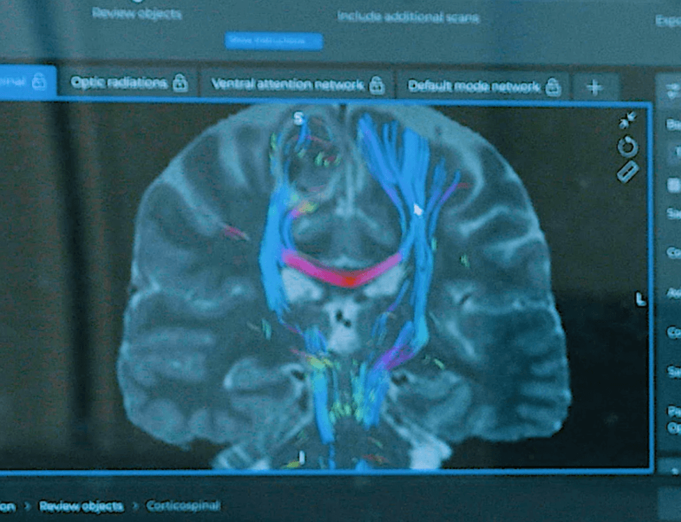
Sensorimotor network and corticospinal tract (CST)

The corticospinal tract (CST) and its importance are well known to surgeons. The corticospinal tract is a group of nerve fibers that carries sensory and motor signals from the brain to the muscles of the body. Damage to the corticospinal tract can result in paralysis or other motor impairments.

In this case, the corticospinal fibers on the right side were visibly disrupted and non-functional, as indicated by the green and red colors on the imaging. The patient had full arm and hand strength, which was reflected visually on the left side, where the fibers were normal.

During the surgery, asleep motor mapping was performed and confirmed the analysis by isolating the functional hand area and non-functional areas on the right side.

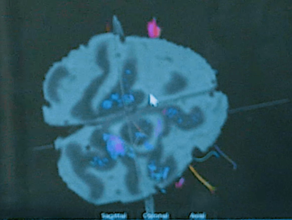
The CST is an important part of the sensorimotor system, which is responsible for sensing physical inputs and controlling voluntary movement. Abnormalities in this network can cause sensory and movement disorders, degenerative diseases, developmental delays and mental health disorders.

image2-1

Networks as seen in Quicktome: Coronal view showing normal corticospinal fibers (right) and non-functional fibers (left)

Surgical decisions and outcomes

Quicktome exports were used in a StealthStation®* surgical navigation platform to reference tractography bundles and network templates. The tumor was accessed on the right side of the brain where there were fewer fibers close to the surface. Post-operative scans analyzed in Quicktome revealed no damage to the fibers and the patient had no resulting deficits.

*StealthStation® is a registered trademark of Medtronic PLC.

image1-1

Three-dimensional view of the tumor

Neurosurgeon

Jonathan Sherman-1
Jonathan Sherman, MD
Neurosurgeon

WVU Medicine, Berkley Medical Center

Rockefeller Neuroscience Institute

Download the case study

case study jonathan sherman 0003

Preserving the corticospinal tract

Download the case study in PDF format.

SEE THE QUICKTOME DIFFERENCE

Upload a free test case, or ask us how Quicktome could work for your site.