Key Highlights:
  • The study of “connectivity”, through magnetic resonance imaging, has a long-standing history in neurosurgery that is backed up by decades of research.
  • Globally, neurosurgeons use connectivity-based solutions every day to provide optimized patient care.
  • Emerging techniques are enhancing the use and removing the barriers to entry for clinical connectivity applications.

Introduction

In the past two decades, connectomics, the study of brain connectivity, has become a mainstay of neuroscience research and has deepened our understanding of the human brain. In light of its recency, it's easy to think of connectivity as a concept that’s still incubating in academia. However, in reality, neurosurgeons have been utilizing connectivity for decades to assess and optimize patient outcomes.

In this article, we present ten papers which demonstrate that connectivity is instrumental to modern neurosurgery. Each manuscript highlights how connectomic analyses are currently implemented in clinical practice and how they are guiding modern surgical approaches with the data provided by diffusion weighted imaging (DWI).

1. Tractography and the Connectome in Neurosurgical Treatment of Gliomas: the Premise, the Progress and the Potential

This paper covers the mechanics, applications, and future of structural connectivity in glioma neurosurgery.

Authors: F. Henderson Jr., K. G. Abdullah, R. Verma, and S. Brem
Publication date: 01/25/2021
Keywords: Connectomics, DTI, diffusion tensor imaging, fiber tractography, glioma surgery


Article 1


At its core, connectivity-based planning helps neurosurgeons determine the location of white matter fiber bundles. From this relatively simple basis, technology has rapidly advanced its practicality in the surgical suite and provided neurosurgeons with new options in pre-surgical planning.

Henderson and colleague’s review emphasize the basic importance of diffusion weighted imaging (DWI) in glioma surgery before describing new modalities in edema correction and tract placement that are being used today. These new modalities are already enhancing patient outcomes by more practically connecting white matter damage to symptoms, determining surgical risk factors, and finding network alterations that serve as biomarkers.


Read more

2. Individualized Map of White Matter Pathways: Connectivity-Based Paradigm for Neurosurgical Planning

This paper introduces a new paradigm which demonstrates that software implementing a connectivity-based clustering method outperforms experts in white matter tract identification and ROI placement.

Authors: B. Tunç, M. Ingalhalikar, D. Parker, J. Lecoeur, N. Singh, R. L. Wolf, L. Macyszyn, S. Brem, and R. Verma
Publication date: 12/16/2015
Keywords: Arcuate fasciculus, diffusion tensor imaging, fractional anistropy, glioma, surgical planning, tractography


Article 2


In Glioma surgery, determining the optimal entry trajectory is one of the most difficult challenges neurosurgeons face. Compounding this challenge is the risk that their chosen trajectory may damage integral eloquent areas of brain tissue. One solution is to manually place Regions of Interest (ROI’s) on the patients pre-surgery brain scan, blocking out eloquent cortical parcels and critical white matter pathways during surgical planning.

Tunc and colleagues however proposed an alternative solution. An imaging algorithm which was trained to identify how the white matter pathways connected to parcels of gray matter, and automatically create ROI’s of eloquent brain tissue surrounding a tumor. Two expert neurosurgeons were consulted and both agreed the algorithm was consistent and superior to manual placement in terms of time saved and ROI’s generated. In a field where time is of the essence, automated solutions are paving the way for optimized patient care.


Read more

3. A Connectomic Atlas of the Human Cerebrum

In this paper, the researchers discuss the Human Connectome Project’s brain atlas, which uses connectivity-based parcellation to map the brain with unprecedented detail.

Authors: C. M. Baker, J. D. Burks, R. G. Briggs, A. K. Conner, C. A. Glenn, G. Sali, T. M. McCoy, J. D. Battiste, D. L. O'Donoghue, M. E. Sughrue
Publication date: 12/01/2018
Keywords: Anatomy, cerebrum, connectivity, DTI, functional connectivity, human, parcellations


Article 3-1


Neuroscience relies on reliability and consistency, particularly when referring to specific parcels of the brain. Naming schemes and parcellation methods vary between sub-specialties, hindering cross-disciplinary efforts.

With the goal of producing a unified schema of the human cortex, Baker and colleagues utilized structural connectivity to define the cortex into eight macroregions, under the nomenclature first introduced by the Human Connectome Project in 2016.

By employing connectivity combined with providing a comprehensive atlas with specific nomenclature, this book provides a framework for patient brain mapping and advocates for accelerating the clinical translation of research.


Read more

4. Surgical Anatomy and Functional Connectivity of the Limbic System

This paper explores the gamut of neuroimaging techniques through the lens of limbic neurosurgery.

Authors: K. Lövblad, K. Schaller
Publication date: 08/27/2009
Keywords: Limbic system, anatomy, magnetic resonance imaging


Article 4


The Limbic system is undoubtedly one of the most integral, yet under-appreciated, systems within the human brain. It is also one of the seven main networks of the human brain. With central roles in motivation, emotional regulation, and memory, damage to this system can have catastrophic consequences for daily life.

Lövblad and Schaller explore the components of the Limbic system, their connections, and the roles they play - with a special focus on how this plays a role in surgical planning and awareness.

Despite coming from the late 2000’s, attention was already focussed on the use of structural connectivity generated through DWI to enable a neurosurgeon to identify damage in, and avoid compromizing, the limbic system before and during surgery.


Read more

5. Brain Functional Magnetic Resonance Imaging Highlights Altered Connections and Functional Networks in Patients With Hypertension

In this paper, Carnevale and colleagues use advanced probabilistic fiber-tracking techniques to discover hypertension-associated microstructural white matter damage.

Authors: L. Carnevale, V. D’Angelosante, A. Landolfi, G. Grillea, G. Selvetella, M. Storto, G. Lembo, D. Carnevale
Publication date: 06/12/2018
Keywords: White matter tractography, hypertension, cognitive impairment, presymptomatic diagnosis


Article 5


Hypertension represents one of the largest surgical risk factors that a patient can face, and in its advanced states, can lead to early-onset dementia and large scale brain damage. The progression of hypertension can go unnoticed for decades, and commonly when identified, the damage has already been done.

In 2018 however, Carnevale and colleagues discovered a new use-case of connectivity analyses: identifying early signs and biomarkers for the onset of hypertensive damage. Structural connectivity analyses revealed abnormalities in diffusion rate along specific white matter tracts, as well as microstructural changes in white matter which corresponded to the degree of cognitive dysfunction and organ damage related to hypertension.

These findings were the first to demonstrate that connectivity can help neurosurgeons identify previously unnoticed neuropathological features that could inform outcome prediction and surgical planning.


Read more

6. Brain Connectomics Applied to Oncological Neuroscience: From a Traditional Surgical Strategy Focusing on Glioma Topography to a Meta-Network Approach

This paper covers meta-networking and the “minimal common brain” and why these may be key to maximizing survival and quality of life in neurooncology.

Authors: H. Duffau
Publication date: 12/22/2020
Keywords: Gliomas, awake mapping, direct electrostimulation, neuroplasticity, connectome, quality of life


Article 6


During glioma surgery, postoperative complications can be tied to changes in brain regions far separated from the location of the tumor. These complications arise from damage caused to large scale brain networks, groups of connected regions in the cortex which can become compromised whilst performing the chosen surgical trajectory.

Duffau and colleagues demonstrate here that connectomic analyses can be utilized during the planning stages of neurosurgery to avoid damage to these connected networks. Moving beyond the idea of “eloquent” and “non-eloquent” brain tissue, they instead argue a model of the “minimal common brain” (MCB), areas of the brain which, if damaged, directly reduce the ability for neuroplasticity and post-surgical recovery. Further, they directly show cases in which the MCB was preserved and compromised, and that in cases where it is preserved, patient survival rates and quality of life improved.


Read more

7. The Parcellation-Based Connectome: Limitations and extensions

This paper explores the impact of map-territory relationships in systems neuroscience.

Authors: M. A. de Reus, M. P. van den Heuvel
Publication date: 10/15/2013
Keywords: Brain network, connectivity, connectome, graph analysis, spatial resolution


Article 7


Metaphorically speaking, the human brain can be thought of as a map of the world, with individual countries - functional gray matter parcels, connected by flight paths - white matter tracts carrying information between them. Functional connectivity analyses measure the rate of communication between two parcels, and map these into a matrix, with each entry on the X- and Y-axes representing an individual parcel.

The process of creating this matrix is better known as “graph theory” and holds key importance for a neurosurgeon attempting to determine which areas may either be compromised by the presence of structural damage, or important to avoid when planning their trajectory.

This review from De Reus and Van Den Heuvel explores graph theory techniques, how to perform them, and their use cases in Neurosurgery. Connectivity-informed surgical planning incorporates modern academic understanding of the cortex, by considering fluidly how parcels are connected and communicating over rigidly defined eloquent and non-eloquent areas.


Read more

8. Diffusion Tensor Imaging of the Corticospinal Tract Before and After Mass Resection as Correlated With Clinical Motor Findings: Preliminary Data

This paper looks back on the history of connectomics with an early preliminary report supporting DTI tractography for neurosurgical planning.

Authors: B. J. Laundre, B. J. Jellison, B. Badie, A. L. Alexander, A. S. Field
Publication date: 07/30/2004
Keywords: Brain network, connectome, connectivity, parcellation, graph analysis, spatial resolution


Article 8


Although the term was yet to be coined, even in 2004 connectomic techniques were being used to tie changes in white matter structure to post surgical improvements.

Laundre and colleagues investigated patients with tumors adjacent to, or invading, the Corticospinal tract (CST). Postoperative motor deficits were only observed in patients where the tumor invaded the CST, and moreso, improved motor recovery was directly related to decreased white matter anisotropy in the CST following surgery.

Although limited in the tools available, this early study laid the groundwork for showing that connectomics had a place in the clinic, even in its infancy.


Read more

9. Connections, Tracts, Fractals, and the Rest: A Working Guide to Network and Connectivity Studies in Neurosurgery

In this paper, you will learn the principles of conducting your own connectivity studies.

Authors: M. G. Hart, R. Romero-Garcia, S. J. Price, T. Santarius, J. Suckling
Publication date: 04/04/2020
Keywords: Connectome, fractal, functional connectivity, structural connectivity, tractography


Article 9


To produce gold standard research, hypotheses must be testable, statistically sound, and most importantly, reproducible by others to enforce their validity. Possibly the greatest roadblock for the translation of academic findings to clinical-use is differences in the methodology and image processing methods employed between academic studies.

With the goal of informing all those interested in connectivity of how to plan out a gold-standard study, Hart and colleagues released this publication as a stand alone guide to conducting connectivity studies with a neurosurgical focus.

The authors ultimately side with Shakespeare, quoting “there is nothing either good nor bad, but thinking makes it so”, suggesting that no study is innately good or bad, but that the design must be dependent on the specific research question being answered. Connectivity is however held in high regard by the authors as a flexible and insightful analytic technique, which is currently and will continue in the future to answer the many mysteries of the brain.


Read more

10. Clinical applications of magnetic resonance imaging based functional and structural connectivity

In this paper, learn how researchers and clinicians are tackling the current limitations of connectivity-based techniques.

Authors: C. Wu, F. Ferreira, M. Fox, N. Harel, J. Hattangadi-Gluth, A. Horn, S. Jbabdi, J. Kahan, A. Oswal, S. A. Sheth, Y. Tie, V. Vakharia, L. Zrinzo, H. Akram
Publication date: 10/11/2021
Keywords: Clinical applications, connectome, connectomics, functional connectivity, structural connectivity, tractography


article 10


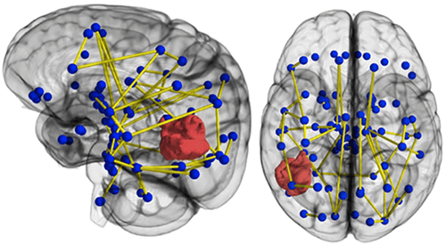
Both structural and functional connectivity analyses have already enhanced our understanding and treatment of several disorders, but they must contend with the variability induced by branching methodologies and human error, and the time required to process large volumes of brain imaging data.

However, novel techniques such as automated machine-learning algorithms are ready to address some of these concerns. By pointing readers to these available solutions, Wu and colleagues review the clinical viability and use cases of connectivity analyses, providing the tactics to confidently put connectivity into practice.


Read more

Conclusion

Collectively, these manuscripts present the case that connectivity-based solutions are an essential component of modern neurosurgery. Their prevalence and the efforts of dedicated clinicians demonstrate that connectivity is easy to implement if you have the right tools. With the right methods, protocols, and knowledge, the future of neurosurgery will be inextricably linked with connectomics. No longer bound to the academics workbench, but instead holding a critical role in ensuring the best possible patient care.