Key Highlights
  • An elderly gentleman who had undergone surgery a year earlier presented with a recurrence of aphasic symptoms
  • Preoperative MRI revealed a large cystic temporal insular glioma with a significant amount of edema
  • Quicktome analysis revealed eloquent tissue underneath the vein of Labbé, and Broca’s area and speech fibers were seen to be located superiorly on top of the tumor
  • Awake motor mapping and switch mapping were conducted, confirming the findings
  • A safe maximal resection (greater than 95%) was achieved with the patient’s speech issues resolving quickly after surgery

Patient history

An elderly gentleman with a recurrent glioblastoma who had undergone surgery a year earlier presented with a recurrence of symptoms. He was becoming aphasic, able to get through approximately 70% of his speech. A preoperative MRI revealed a large cystic temporal insular glioma with a significant amount of edema. The tumor was IDH-wild type, MGMT negative.

Quicktome analysis revealing fibers of connecting speech areas around the tumor.

Quicktome analysis revealing fibers of connecting speech areas around the tumor.

Brain network involvement

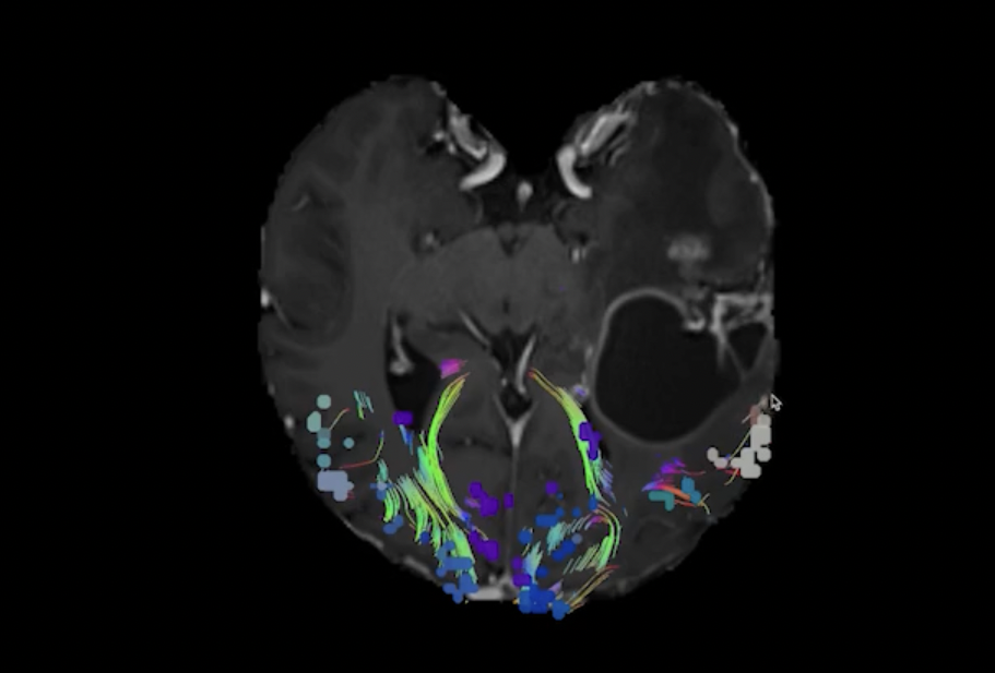
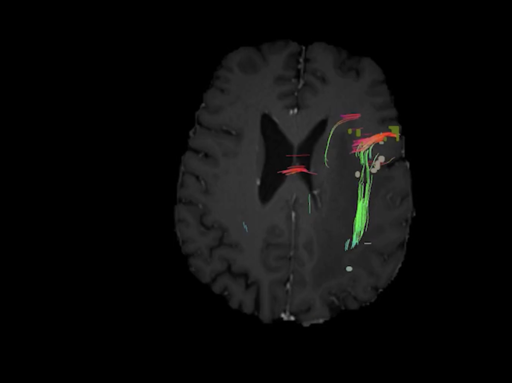
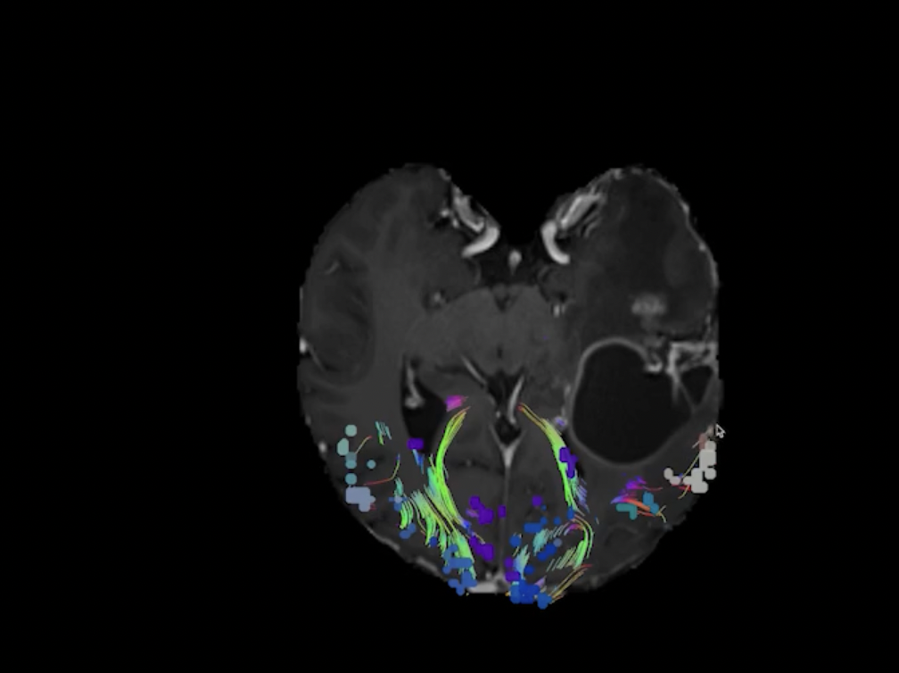
The patient would be typically pre-treated with Decadron and Mannitol to optimize brain swelling, but with Quicktome, the tractography and the connections in the brain were clearly visible, specifically the language corridor.

Quicktome analysis revealed that Broca’s area and the fibers connecting the speech areas were located superiorly on top of the tumor. The overhanging temporal lobe brain on the left, underneath the vein of Labbé, was shown to be eloquent tissue.

Quicktome analysis showed that everything interior to the tumor could be resected, but deep and posterior the boundary of the cyst needed to be respected. The anterior temporal lobe was deemed somewhat non-functional and therefore a safe area to resect.

The surgeon was able to reassure the patient and their family about the risks, the anatomy involved and expected outcomes.

Brain networks as seen in Quicktome: eloquent tissue revealed underneath the vein of Labbé.

Brain networks as seen in Quicktome: eloquent tissue revealed underneath the vein of Labbé.

Surgical decisions and outcomes

An approach was taken through the anterior temporal lobe followed by a posterior trajectory. Both awake motor mapping and switch mapping were conducted, and the findings coincided with the findings of Quicktome prior to the operation. The overlying temporal lobe of the overlying cyst was confirmed to be an area of Warnicke’s, very critical to the patient’s speech center. A safe maximal resection of the tumor (greater than 95%) was achieved, with postoperative scans revealing minimal contrast enhancement. The patient's speech issues resolved quickly after surgery.

Download the case study

CAS0005 Image

Recurrent glioblastoma and restoring speech function

Download the case study in PDF format.

SEE THE QUICKTOME DIFFERENCE

Upload a free test case, or ask us how Quicktome could work for your site.